. 2008 Jan 23;27(2):373-83.
doi: 10.1038/sj.emboj.7601962. Epub 2007 Dec 13.

A critical role of RICK/RIP2 polyubiquitination in Nod-induced NF-kappaB activation

Affiliations

A critical role of RICK/RIP2 polyubiquitination in Nod-induced NF-kappaB activation

Mizuho Hasegawa et al. EMBO J. .

Abstract

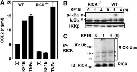
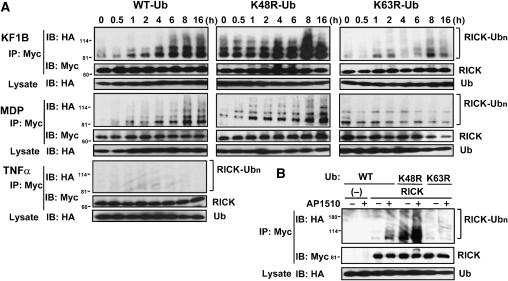
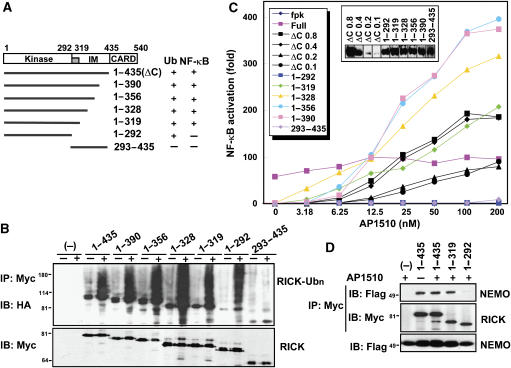
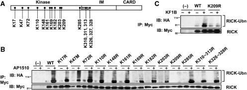
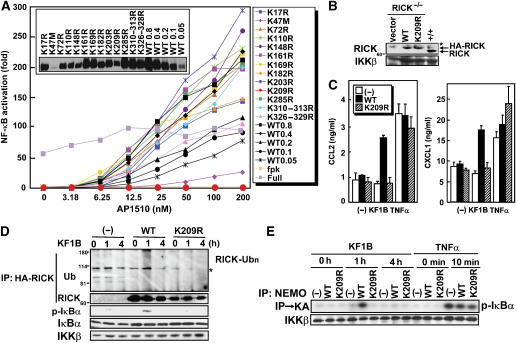
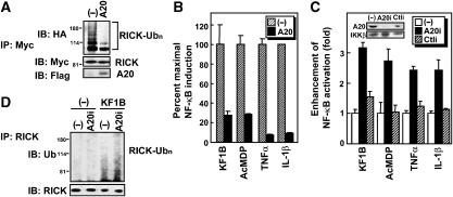
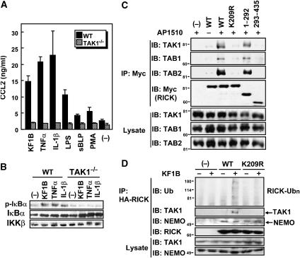
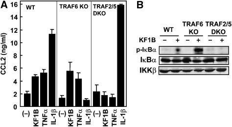
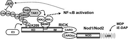
Nod1 and Nod2 are intracellular proteins that are involved in host recognition of specific bacterial molecules and are genetically associated with several inflammatory diseases. Nod1 and Nod2 stimulation activates NF-kappaB through RICK, a caspase-recruitment domain-containing kinase. However, the mechanism by which RICK activates NF-kappaB in response to Nod1 and Nod2 stimulation is unknown. Here we show that RICK is conjugated with lysine-63-linked polyubiquitin chains at lysine 209 (K209) located in its kinase domain upon Nod1 or Nod2 stimulation and by induced oligomerization of RICK. Polyubiquitination of RICK at K209 was essential for RICK-mediated IKK activation and cytokine/chemokine secretion. However, RICK polyubiquitination did not require the kinase activity of RICK or alter the interaction of RICK with NEMO, a regulatory subunit of IkappaB kinase (IKK). Instead, polyubiquitination of RICK was found to mediate the recruitment of TAK1, a kinase that was found to be essential for Nod1-induced signaling. Thus, RICK polyubiquitination links TAK1 to IKK complexes, a critical step in Nod1/Nod2-mediated NF-kappaB activation.


Figures

Figure 1
Figure 1
Nod1 stimulation induces ubiquitination of RICK. (A) MEFs from WT and RICK-deficient mice were stimulated with 5 μg/ml KF1B (synthetic Nod1-stimulatory compound), 10 ng/ml TNFα or left alone. Twenty-four hours post-stimulation, the levels of CCL2 in culture supernatant were determined by ELISA. The results shown are given as mean±s.d. of triplicate cultures and are representative of three experiments. (B) WT or RICK-deficient MEFs were stimulated with 5 μg/ml KF1B for the indicated times. Post-stimulation, cells were lysed and immunoblotted with anti-phospho-IκBα, anti-IκBα or IKKβ Ab. (C) MEFs were stimulated with or without KF1B for the indicated times. Post-stimulation, cells were lysed and RICK was immunoprecipitated with rabbit anti-RICK Ab. Ubiquitinated (upper panel) and total (lower panel). RICK proteins in the immunoprecipitates were immunodetected by mouse monoclonal anti-RICK and anti-Ub Abs, respectively. Nonspecific signals detected with the Ab are indicated by asterisks.
Figure 2
Figure 2
Nod1 and Nod2 stimulation induce K63-linked polyubiquitination of RICK. (A) HEK293 cells constitutively expressing Nod1 (for KF1B stimulation), Nod2 (for MDP stimulation) and parental HEK293 cells (for TNFα stimulation) were transfected with expression plasmid of Myc-RICK in the presence of expression plasmids of HA-tagged WT, K48R, K63R mutant Ub proteins. Cells were stimulated with 100 ng/ml KF1B, 100 ng/ml MDP (synthetic Nod2-stimulatory compound) or 10 ng/ml TNFα, or left alone (0 h control) for the indicated times. Myc-RICK proteins in transfected cells were immunoprecipitated with rabbit anti-Myc Ab and ubiquitinated RICK proteins were then detected by immunoblotting with anti-HA Ab (upper panels labeled as RICK-Ubn). As a control, RICK proteins in the same samples and Ub proteins in total lysate were detected by immunoblotting with the indicated Abs. (B) Oligomerization-dependent RICK ubiquitination. HEK293T cells were transfected with control pcDNA3-RICK-Fpk3-Myc (−) or pcDNA3-RICK-Fpk3-Myc (RICK) in the presence of HA-Ub expression plasmids. Eighteen hours post-transfection, cells were treated with or without 200 nM AP1510. Twenty-four hours post-transfection, proteins were immunoprecipitated and ubiquitinated RICK was analyzed as described in panel A.
Figure 3
Figure 3
The kinase domain of RICK is essential for RICK ubiquitination, but is not sufficient to activate NF-κB. (A) Schematic diagram of the structural domains of RICK and different deletion mutants. The results of ubiquitination and NF-κB activation experiments are summarized on the right. The region, which requires NF-κB activation in the IM domain is indicated by a gray box. (B) HEK293T cells were transfected with pcDNA3-RICK-(1–435)-Fpk3-Myc (ΔC), RICK-(1–292)-Fpk3-Myc, RICK-(1–319)-Fpk3-Myc, RICK-(1–328)-Fpk3-Myc, RICK-(1–356)-Fpk3-Myc, RICK-(1–390)-Fpk3-Myc RICK-(293–435)-Fpk3-Myc and control vector in the presence of HA-Ub expression plasmid and treated with AP1510 as in Figure 2B. Twenty-four hours post-transfection, proteins were immunoprecipitated and ubiquitinated RICK were analyzed as described in Figure 2. (C) HEK293T cells were transfected with pcDNA3-RICK-(1–435)-Fpk3-Myc (ΔC), RICK-(1–292)-Fpk3-Myc, RICK-(1–319)-Fpk3-Myc, RICK-(1–328)-Fpk3-Myc, RICK-(1–356)-Fpk3-Myc, RICK-(1–390)-Fpk3-Myc RICK-(293–435)-Fpk3-Myc and control vector in the presence of reporter plasmids. Eight hours post-transfection, cells were treated with medium containing the indicated amounts of AP1510. Twenty-four hours post-transfection, ligand-dependent NF-κB activation was determined with reporter assay. The level of NF-κB-dependent transcription activity in cells transfected with control vector is given as 1. The expression levels of Myc-RICK proteins are shown in an inset. (D) HEK293T cells were transfected with control vector (−), pcDNA3-RICK-(1–435)-Fpk3-Myc, RICK-(1–319)-Fpk3-Myc and RICK-(1–292)-Fpk3-Myc in the presence of reporter plasmids, and treated with AP1510 as in Figure 2B. Twenty-four hours post-transfection, RICK proteins were immunoprecipitated with rabbit polyclonal anti-Myc Ab, and then Flag-NEMO (upper panel) and Myc-RICK (middle panel) were immunodetected with mouse monoclonal anti-Flag and anti-Myc Abs, respectively. As control, Flag-NEMO in total lysate was immunodetected with anti-Flag Ab and shown in the lower panel.
Figure 4
Figure 4
Lysine 209 is ubiquitinated in the kinase domain of RICK. (A) Location of lysine residues, which were replaced by arginines in RICK site-directed mutants. (B) HEK293T cells were transfected with control vector (−), pcDNA3-RICK-ΔCARD-Fpk3-Myc (WT) and various lysine-to-arginine mutants in the presence HA-Ub expression plasmid and treated with AP1510 as in Figure 2B. Twenty-four hours post-transfection, proteins were immunoprecipitated and ubiquitinated RICK were immunodetected as described in Figure 1. (C) HEK293 cells constitutively expressing Nod1 were transfected with the expression plasmids of WT and K209R-mutant Myc-RICK in the presence of HA-Ub expression plasmid. Eighteen hours post-transfection, cells were stimulated with or without 100 ng/ml KF1B. Twenty-four hours post-transfection, proteins were immunoprecipitated and ubiquitinated RICK were immunodetected as described in Figure 1.
Figure 5
Figure 5
Polyubiqutination at K209 is essential for RICK-mediated signaling. (A) HEK293T cells were transfected with pcDNA3-RICK-ΔCARD-Fpk3-Myc (WT) or the mutants in the presence of reporter plasmids. Eight hours post-transfection, cells were treated with medium containing the indicated amount of AP1510. Twenty-four hours post-transfection, ligand-dependent NF-κB activation was determined with reporter assay. The level of NF-κB-dependent transcription activity in cells transfected with control vector is given as 1. Myc-tagged RICK proteins detected with anti-Myc Ab are shown in the inset. (B) RICK-deficient MEFs were infected with retrovirus vectors expressing WT or K209R HA-RICK or control vector and selected by hygromycine. RICK proteins detected with anti-RICK Ab. A nonspecific signal detected with the Ab is indicated by an asterisk. (C) RICK-deficient MEFs were infected with control (−) or retrovirus vectors expressing WT or K209R HA-RICK. Infected MEFs were selected by hygromycin and stimulated with 5 μg/ml KF1B, 10 ng/ml TNFα or left alone. Twelve hours post-stimulation, secretion levels of CCL2 and CXCL1 were determined by ELISA. The results shown are given as mean±s.d. of triplicate cultures and are representative of three experiments. (D) MEFs stably expressing RICK or RICK K209R were stimulated with 5 μg/ml KF1B for the indicated time. Post-stimulation, cells were lysed, RICK was immunoprecipitated and ubiquitinated RICK was immunodetected by anti-Ub Ab (upper panel). Cell lysate was also immunoblotted with anti-phospho-IκBα, anti-IκBα or IKKβ Ab (bottom three panels). A nonspecific signal detected with the Ab is indicated by an asterisk. (E) MEFs stably expressing RICK or RICK K209R were stimulated with 5 μg/ml KF1B or TNFα for indicated times, and then anti-NEMO Ab was used to immunoprecipitate the IKK complex. The IKK assay was carried out by using GST-IκBα and γ-32P-ATP as substrates (upper). The amount of IKKβ in the immunoprecipitates was detected by immunoblotting.
Figure 6
Figure 6
A20 functions as a negative regulator of Nod1 and Nod2 signaling via deubiquitination of RICK. (A) HEK293T cells were transfected with Flag-tagged A20 expression plasmids or control vector (−) in the presence of pcDNA3-RICK-ΔCARD-Fpk3-Myc (WT) and HA-Ub expression plasmid. Eighteen hours post-transfection, cells were treated with 200 nM AP1510 or left alone. Twenty-four hours post-transfection, proteins immunoprecipitated with rabbit anti-Myc Ab were subjected to SDS–PAGE and analyzed by western blot analysis using anti-HA, Myc and Flag Abs to detect ubiquitinated RICK proteins, RICK proteins, and A20 protein, respectively. (B) HEK293T cells were transfected with expression plasmids of A20 or control vector in the presence of reporter plasmids. Eight post-transfection, cells were treated with medium containing 1 μg/ml KF1B, 1 μg/ml AcMDP, 10 ng/ml TNF or 10 ng/ml IL-1β. Twenty-four hours post-transfection, ligand-dependent NF-κB activation was determined with reporter assay. The level of NF-κB-dependent transcription activity in the absence of A20 is given as 100%. The results shown are given as mean±s.d. of triplicate cultures and are representative of three experiments. (C) HEK293T cells were transfected with pcDNA3 (−), A20 interference RNA (RNAi) plasmid or control RNAi plasmid (Ctli) in the presence of reporter plasmids. Thirty-six hours after transfection, cells were treated with medium containing 1 μg/ml KF1B, 1 μg/ml AcMDP, 10 ng/ml TNFα or IL-1β. Forty-eight hours post-transfection, ligand-dependent NF-κB activation was determined with reporter assay. The level of NF-κB-dependent transcription activity in cells transfected with pcDNA3 is given as 1. Expression of A20 and control IKKβ proteins is shown in inset. The results shown are given as mean±s.d. of triplicate cultures and are representative of three experiments. (D) HEK293 cells constitutively expressing Nod1 were transfected with pcDNA3 (−) or A20 RNAi plasmid. Forty-eight hours after transfection, cells were treated with 100 ng/ml KF1B for 1 h. The cells were lysed and endogenous RICK was immunoprecipitated. Ubiquitinated and total RICK proteins were immunodetected with anti-Ub and anti-RICK Abs, respectively.
Figure 7
Figure 7
RICK polyubiquitination at K209 is required for the recruitment of TAK1/TAB1/TAB2 complex. (A) WT or TAK1-deficient MEFs were stimulated with 5 μg/ml KF1B, 10 ng/ml TNFα, 10 ng/ml IL-1β, 100 ng/ml LPS, 1 μg/ml sBLP, 50 ng/ml PMA plus 0.7 μg/ml Ca2+ ionophore A23187 or left alone. Twenty-four hours post-stimulation, secretion levels of CCL2 were determined by ELISA. The results shown are given as mean±s.d. of triplicate cultures and are representative of three experiments. (B) WT or TAK1-deficient MEFs were stimulated with 5 μg/ml KF1B, 10 ng/ml TNFα or 10 ng/ml IL-1β for 1 h. Post-stimulation, cells were lysed and lysates immunoblotted with anti-phospho-IκBα, anti-IκBα or IKKβ Ab. (C) HEK293T cells were transfected with expression plasmids of RICK-ΔCARD-Fpk3-Myc (WT), RICK-K209R-ΔCARD-Fpk3-Myc (K209R), RICK-(1–292)-Fpk3-Myc, RICK-(293–435)-Fpk3-Myc and control vector (−) in the presence of expression plasmids of Ub, HA-TAK1, Flag-TAB1 and T7-TAB2 and treated with AP1510 as in Figure 2B. Twenty-four hours post-transfection, proteins immunoprecipitated with rabbit anti-Myc Ab were subjected to SDS–PAGE and analyzed by immunoblotting analysis using anti-HA or, anti-Flag, anti-T7 and anti-Ub Abs. (D) MEFs stably expressing RICK or RICK K209R were stimulated with 5 μg/ml KF1B for 1 h. The cells were lysed, RICK was immunoprecipitated and endogenous Ub, TAK1, NEMO and RICK were immunodetected with anti-Ub, anti-TAK1 Ab, anti-NEMO or anti-RICK antibodies.
Figure 8
Figure 8
TRAF2/TRAF5, but not TRAF6, are required for Nod1-mediated NF-κB activation and CCL2 secretion. (A) WT, TRAF6−/− (TRAF6 KO) or TRAF2−/−TRAF5−/− (TRAF2/5 DKO) MEFs were stimulated with 5 μg/ml KF1B, 10 ng/ml TNFα, 10 ng/ml IL-1β or left alone. After 24 h incubation, the secretion levels of CCL2 were determined by ELISA. The results shown are given as mean±s.d. of triplicate cultures and are representative of three experiments. (B) WT and mutant MEFs were stimulated with 5 μg/ml KF1B for 1 h or left alone. The phosphorylated (upper panel) and whole (middle panel) populations of IκBα and IKKβ (lower panel) were immunodetected with specific Abs.
Figure 9
Figure 9
Model for the role of RICK polyubiquitination in Nod signaling. For detail, see text.

References

    1. Abbott DW, Wilkins A, Asara JM, Cantley LC (2004) The Crohn's disease protein, NOD2, requires RIP2 in order to induce ubiquitinylation of a novel site on NEMO. Curr Biol 14: 2217–2227 - PubMed
    1. Abbott DW, Yang Y, Hutti JE, Madhavarapu S, Kelliher MA, Cantley LC (2007) Coordinated regulation of Toll-like receptor and NOD2 signaling by K63-linked polyubiquitin chains. Mol Cell Biol 27: 6012–6025 - PMC - PubMed
    1. Boone DL, Turer EE, Lee EG, Ahmad RC, Wheeler MT, Tsui C, Hurley P, Chien M, Chai S, Hitotsumatsu O, McNally E, Pickart C, Ma A (2004) The ubiquitin-modifying enzyme A20 is required for termination of Toll-like receptor responses. Nat Immunol 5: 1052–1060 - PubMed
    1. Boughan PK, Argent RH, Body-Malapel M, Park JH, Ewings KE, Bowie AG, Ong SJ, Cook SJ, Sorensen OE, Manzo BA, Inohara N, Klein NJ, Nunez G, Atherton JC, Bajaj-Elliott M (2006) Nucleotide-binding oligomerization domain-1 and epidermal growth factor receptor: critical regulators of beta-defensins during Helicobacter pylori infection. J Biol Chem 281: 11637–11648 - PubMed
    1. Chamaillard M, Hashimoto M, Horie Y, Masumoto J, Qiu S, Saab L, Ogura Y, Kawasaki A, Fukase K, Kusumoto S, Valvano MA, Foster SJ, Mak TW, Nunez G, Inohara N (2003) An essential role for NOD1 in host recognition of bacterial peptidoglycan containing diaminopimelic acid. Nat Immunol 4: 702–707 - PubMed

Publication types

MeSH terms

Substances