. 2008 Jan 9;27(1):122-31.
doi: 10.1038/sj.emboj.7601957. Epub 2007 Dec 13.

Intrasplicing coordinates alternative first exons with alternative splicing in the protein 4.1R gene

Affiliations

Intrasplicing coordinates alternative first exons with alternative splicing in the protein 4.1R gene

Marilyn K Parra et al. EMBO J. .

Abstract

In the protein 4.1R gene, alternative first exons splice differentially to alternative 3' splice sites far downstream in exon 2'/2 (E2'/2). We describe a novel intrasplicing mechanism by which exon 1A (E1A) splices exclusively to the distal E2'/2 acceptor via two nested splicing reactions regulated by novel properties of exon 1B (E1B). E1B behaves as an exon in the first step, using its consensus 5' donor to splice to the proximal E2'/2 acceptor. A long region of downstream intron is excised, juxtaposing E1B with E2'/2 to generate a new composite acceptor containing the E1B branchpoint/pyrimidine tract and E2 distal 3' AG-dinucleotide. Next, the upstream E1A splices over E1B to this distal acceptor, excising the remaining intron plus E1B and E2' to form mature E1A/E2 product. We mapped branchpoints for both intrasplicing reactions and demonstrated that mutation of the E1B 5' splice site or branchpoint abrogates intrasplicing. In the 4.1R gene, intrasplicing ultimately determines N-terminal protein structure and function. More generally, intrasplicing represents a new mechanism by which alternative promoters can be coordinated with downstream alternative splicing.


Figures

Figure 1
Figure 1
Promoter choice and downstream alternative splicing events in protein 4.1R. (A) Arrangement of the 4.1R gene, 5′ region, showing three alternative first exons E1A, E1B, and E1C, and the differential manner in which they splice selectively to alternative acceptors downstream in E2. The proximal site E2 acceptor is pyrimidine rich and is predicted to be a stronger 3′ splice site than the distal, more purine-rich site (Tan et al, 2005); the 17 bp region between these acceptors is indicated by E2′. The thin line to E1B indicates that this isoform is rare among database clones. Numbers on the introns indicate length in kilobases. (B) E2′ (shaded) and its flanking sequence. The branchpoint (BP) and alternative 3′ splice acceptor sites are indicated. Translation start codon in E2′ is underlined. (C) An important functional consequence of this splicing variation is that E1A transcripts produce a shorter protein isoform compared with that made from E1B/C promoters, with the former missing a critical ‘headpiece' region (HP) that participates in protein–protein interactions. Other protein 4.1R domains are indicated by the abbreviations FERM (4.1/ezrin/radixin/moesin homology domain), SAB (spectrin-actin binding domain), and CTD (C-terminal domain).
Figure 2
Figure 2
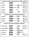
Minigene constructs used to analyze intrasplicing. Diagrams show the structure of minigenes used in this study to map the intrasplicing regulatory element, to test promoter dependence of intrasplicing, and to investigate motifs essential for the intrasplicing mechanism. Each construct consists of three components derived from the human 4.1R gene: a promoter/first exon, a portion of the 4.1R intron downstream of E1A, and a terminal exon consisting of 4.1R E2 with its proximal upstream intron sequence. Gaps represent regions of the natural gene that have been deleted. Promoter identity is indicated in the construct designation; pE1A, 4.1R E1A promoter; pCMV, CMV promoter; pANK, erythroid ANK1 promoter (Gallagher et al, 2000); and pE1C, 4.1R E1C promoter. E1B does not have a consensus 3′ splice site; its approximate 5′ boundary is indicated by the dotted line. Constructs pE1A-2b and pE1A-2a each contains only half of the E1B region. In the bottom panel, sites marked by the ‘X' indicate position of mutations in the E1B 5′ splice site and E1B-associated branchpoint; 1B* indicates a candidate regulatory element from the paralogous 4.1B gene.
Figure 3
Figure 3
A downstream regulatory element is required for proper E1A splicing to E2. Analysis of spliced products as a function of the presence of downstream regulatory element(s). Shown are the amplified products from cells transfected with the following constructs: lane 1, size standards (indicated in nt); lane 2, pE1A-1; lane 3, pE1A-2L; lane 4, pE1A-2; lane 5, pE1A-2a; lane 6, pE1A-2b. Lane 7 represents a negative PCR control. The larger PCR product (expected size: 343 nt) corresponds to inappropriate splicing of E1A to the proximal acceptor in E2, whereas the smaller product (expected size: 326 nt) represents correct splicing to the distal acceptor. Correct splicing of transcripts initiated at E1A required a regulatory element in the downstream intron that overlaps with E1B.
Figure 4
Figure 4
Proper E1A splicing is independent of promoter architecture, but requires splicing element(s) associated with E1B. (A) Analysis of spliced products as a function of promoter identity. Shown are the amplified products from cells transfected with the following constructs: lanes 1 and 7, pE1A-2L; lane 2, pCMV-2L; lane 3, pANK-2L; lanes 4 and 6, pE1A-1 negative control, lanes 5 and 9, mock transfection; lane 8, pE1C-2L. Correct splicing of transcripts initiated at promoters upstream of E1B was independent of promoter or first exon identity. (B) Dependence of E1A splicing on the E1B 5′ splice site. Lanes in (B) are as follows: lane 1, construct E1A-1 lacking the regulatory element; lane 2, construct E1A-2 with an intact E1B 5′ splice site; lane 3, construct E1A-2mut5′ with a mutant E1B 5′ splice site; lane 4, negative PCR control; lane 5, mock-transfected cells.
Figure 5
Figure 5
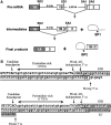
The two-step intrasplicing hypothesis. (A) In this model, the splice donor site of E1B (SD1) splices to the proximal 3′ acceptor of E2 (SA1) in the first step of the reaction, resulting in an intermediate and an excised lariat structure. This step juxtaposes E1B with E2 and, by eliminating SA1, activates distal acceptor SA2. In the second step, E1B functions as an intron so that the E1A 5′ splice site (SD2) splices to SA2 using branchpoint bp2, excising a second lariat and releasing the mature product. (B) Top panel shows the sequence context of E1B prior to the first step of intrasplicing, when it is preceded by a candidate branchpoint/pyrimidine tract and followed by a consensus 5′ splice site. Lower panel shows the sequence context after the first step of intrasplicing, when E1B has been juxtaposed to E2′/2 to form a new composite 3′ splice site in conjunction with the distal AG-dinucleotide.
Figure 6
Figure 6
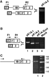
Analysis of the partially spliced RNA intermediate. (A) Detection of the intermediate RNA produced in the first step of intrasplicing from pE1A-2. RNA from transfected cells was amplified by RT–PCR using the indicated primers. (B) Splicing of intermediate RNA into mature product. Constructs used for splicing assays: lane 1, pE1A-1 negative control; lane 2, pE1A-2L positive control; lane 3, intermediate pE1A-2Lint; lane 4, mock-transfected control. Note that the intermediate functions as a direct precursor of the mature E1A-E2 spliced product. (C) Detection of a partial splicing intermediate from endogenous RNA. Natural RNA purified from mouse splenic erythroblasts was amplified using an antisense primer at the E2′/2 junction, and sense primers in the intron upstream of E1B. In the longest successful amplification (lane 1), approximately 3.2 kb of upstream intron was present upstream of the joined E1B/2′/2. Lane 2 represents size standards in kilobases.
Figure 7
Figure 7
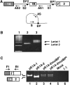
Branchpoint analysis. (A) RT–PCR strategy for detecting lariat intermediates from both steps of intrasplicing (Vogel et al, 1997), showing location of antisense (AS1, AS2) and sense (S1, S2) primers. Branchpoint is indicated by ‘BP'. (B) Gel analysis of the amplified branchpoint structures. Arrows indicate PCR products whose sequences were confirmed to represent lariat structures in which upstream 5′ splice sites loop back to the relevant branchpoints for both steps of the proposed mechanism. Lane 1, 100 bp size ladder; lane 2, lariat from second step of intrasplicing; lane 3, lariat from first step of intrasplicing. The additional band in lane 3 is an artifact unrelated to 4.1R transcripts. (C) Functional testing of the branchpoint for step 2. Lane 1, incorrectly spliced product from pE1A-1; lane 2, correctly spliced product from pE1A-2; lanes 3–4, mixture of spliced products from two different branchpoint mutants; lane 5, mock transfection; lane 6, negative PCR control. Mutagenesis of the highly conserved branchpoint resulted in reduced production of properly spliced E1A transcript.
Figure 8
Figure 8
Phylogenetic conservation of the intraexon-associated branchpoint and 5′ splice site. (Above) Organization of the intrasplicing element, showing the conserved branchpoint located 84–92 nt upstream of the conserved, consensus 5′ splice site. (Below) Comparison of genome sequences shows that these two key motifs were conserved in a wide range of mammalian species. In the branchpoint sequences, nonconserved nucleotides are underlined and the presumed branchpoint A is represented in bold. In the 5′ splice site sequences, upper case represents the end of E1B and lower case nucleotides indicate the beginning of the intron.
Figure 9
Figure 9
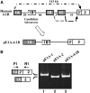
Functional analysis of a heterologous intraexon from the paralogous 4.1B gene. (A) A candidate intraexon E1B* identified 20 kb downstream of E1A in the 4.1B gene was inserted into the 4.1R minigene in place of the natural element, and tested for its ability to promote the intrasplicing pathway. (B) Splicing assay showing that the heterologous element does promote proper intrasplicing.

References

    1. Akerman M, Mandel-Gutfreund Y (2006) Alternative splicing regulation at tandem 3′ splice sites. Nucleic Acids Res 34: 23–31 - PMC - PubMed
    1. Auboeuf D, Dowhan DH, Kang YK, Larkin K, Lee JW, Berget SM, O'Malley BW (2004) Differential recruitment of nuclear receptor coactivators may determine alternative RNA splice site choice in target genes. Proc Natl Acad Sci USA 101: 2270–2274 - PMC - PubMed
    1. Baek D, Davis C, Ewing B, Gordon D, Green P (2007) Characterization and predictive discovery of evolutionarily conserved mammalian alternative promoters. Genome Res 17: 145–155 - PMC - PubMed
    1. Baklouti F, Huang SC, Vulliamy TJ, Delaunay J, Benz EJ Jr (1997) Organization of the human protein 4.1 genomic locus: new insights into the tissue-specific alternative splicing of the pre-mRNA. Genomics 39: 289–302 - PubMed
    1. Bird G, Zorio DA, Bentley DL (2004) RNA polymerase II carboxy-terminal domain phosphorylation is required for cotranscriptional pre-mRNA splicing and 3′-end formation. Mol Cell Biol 24: 8963–8969 - PMC - PubMed

Publication types

MeSH terms