. 2008 Jan 9;27(1):155-67.
doi: 10.1038/sj.emboj.7601948. Epub 2007 Dec 13.

Sequential recruitment of the repair factors during NER: the role of XPG in initiating the resynthesis step

Affiliations

Sequential recruitment of the repair factors during NER: the role of XPG in initiating the resynthesis step

Vincent Mocquet et al. EMBO J. .

Abstract

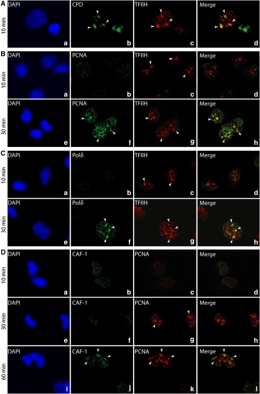
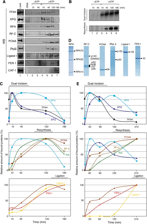
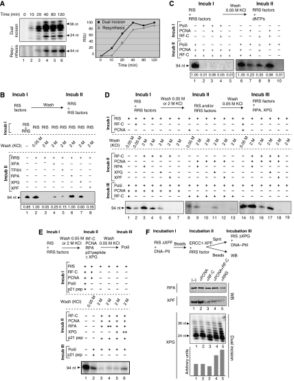
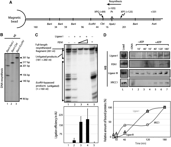
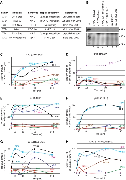
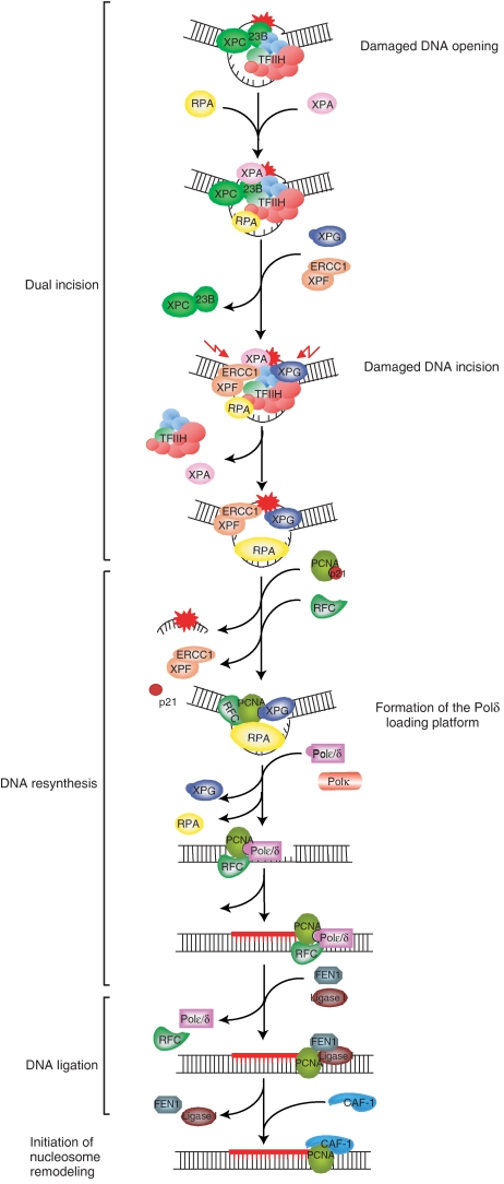
To address the biochemical mechanisms underlying the coordination between the various proteins required for nucleotide excision repair (NER), we employed the immobilized template system. Using either wild-type or mutated recombinant proteins, we identified the factors involved in the NER process and showed the sequential comings and goings of these factors to the immobilized damaged DNA. Firstly, we found that PCNA and RF-C arrival requires XPF 5' incision. Moreover, the positioning of RF-C is facilitated by RPA and induces XPF release. Concomitantly, XPG leads to PCNA recruitment and stabilization. Our data strongly suggest that this interaction with XPG protects PCNA and Pol delta from the effect of inhibitors such as p21. XPG and RPA are released as soon as Pol delta is recruited by the RF-C/PCNA complex. Finally, a ligation system composed of FEN1 and Ligase I can be recruited to fully restore the DNA. In addition, using XP or trichothiodystrophy patient-derived cell extracts, we were able to diagnose the biochemical defect that may prove to be important for therapeutic purposes.


Figures

Figure 1
Figure 1
In vivo sequential recruitment of NER factors. Rescued XPCS2BA human fibroblasts were locally UV irradiated and labelled at 10, 30 and 60 min after UV irradiation with the indicated MAbs or PAbs. Colocalization of (A) CPD and TFIIH (XPB) (panels a–d), (B) TFIIH and PCNA (panels a–h), (C) TFIIH and Polδ (panels a–h) and (D) PCNA and CAF1 (panels a–l). Nuclei were counterstained with DAPI, and pictures were merged.
Figure 2
Figure 2
In vitro sequential recruitment of the NER factors. (A) The immobilized damaged DNA fragment was incubated with NE. At different time points, the immobilized DNA was washed with 0.05 M KCl and the remaining bound factors were further analysed by western blot. (B) The damaged fragment removal (Dual Incision) and the gap filling (Resynthesis) activities were also followed through time (Supplementary data 1). (C) The WB signals were quantified using Genetool (Syngene) and plotted on the graphs as a percentage of the maximal binding to the DNA. (D) Coomassie staining of the highly purified human NER resynthesis factors RPA, RF-C, PCNA, Polδ, Ligase I and FEN 1. (E) The same recruitment experiment as in (B) was carried out with our complete reconstituted system (dual incision, resynthesis and ligation factors). All these experiments were carried out at least two times.
Figure 3
Figure 3
Molecular events during the resynthesis. (A) Time course of the dual incision (upper panel) and the resynthesis (middle panel). Signals were quantified (Genetool, Syngene) and plotted in a graph (square for dual incision; triangle for DNA resynthesis) (lower panel). (B) The immobilized DNA-Pt was incubated with RIS+/−RRS (Incub I). After washes at either 0.05 M KCl or 2 M KCl, DNA resynthesis were performed using RRS supplemented with some of the RIS factors as indicated (Incub II). The relative intensity of each signal is indicated at the bottom of the gel. (C) DNA-Pt was incubated with RIS either alone or in combination with the indicated RRS factors (Incub I). After 0.05 M KCl wash, the complementary RRS factors were added for DNA resynthesis (Incub II). The relative intensity of each signal is indicated at the bottom of the gel. (D) DNA-Pt was incubated with RIS either alone or with the indicated RRS factors (Incub I). After washes with either 0.05 M KCl or 2 M KCl, Pt-DNA was further incubated with the indicated RIS and/or the RRS factors (Incub II). Following a 0.05 M KCl wash, and the addition of the complementary RRS factors (Incub III), DNA resynthesis was checked. (E) The experiment was carried out similarly as described in (D), except that we added 2 nmol of a peptide corresponding to the domain of interaction between the p21 CDK inhibitor and PCNA as indicated. (F) RIS-ΔXPF and DNA-Pt were incubated with the RRS factors as indicated at the top of the panels. Following Incub I+Incub II, and a 0.05 M KCl wash, amounts of RPA (upper panel) and XPF (middle panel) remaining on the DNA fragment were checked by western blot. The released XPG (lower panel) was tested in dual incision with RIS-ΔXPG, containing DNA-PtII as a challenge template (Riedl et al, 2003). A graph depicts the relative intensity of each signal.
Figure 4
Figure 4
The ligation in NER. (A) Scheme of the substrate used with the position of the lesion, the restriction sites and the endonucleases cut sites. The sense of resynthesis is indicated. (B) DNA-Pt was incubated with a NE. The repaired DNA was then digested with indicated restriction enzymes. The absence of the 96 nt signal proved that the ligation occurred between ClaI and EcoRV (lane3). (C) The ligation activity was investigated by incubating the DNA with the RIS+RRS and combinations of the ligation system (RLS) before digestion by EcoRV and BanI. The unligated DNA due to the absence of ligation could be easily discriminated from the unligated DNA due to nick translation process, bypassing the EcoRV site. To evaluate the ligation efficiency, the ratio between the full-length resynthesized DNA and all the forms of unligated DNA was calculated. (D) The recruitment of Ligase III and XRCC1 on the damaged DNA was investigated with a method described in Figure 2. Western blot (WB) signals (upper panel) were quantified and plotted (lower panel) (open circle: XRCC1; open triangle: Ligase III).
Figure 5
Figure 5
Recruitment of the NER machinery involving mutated factors. (A) Data concerning the mutated factors used and references. (B) Dual incision and resynthesis assays were carried out with each mutant (lanes 2–8). WT factors were used for the positive control (lane 1). The recruitment analysis of the NER factors was carried out as described previously with mutated forms of (C) XPC, (E) XPB and (G) XPA used in a reconstituted system and (D) XPD, (F) p8 and (H) XPG coming from patient cell extracts.
Figure 6
Figure 6
Sequential complexes in NER from the damage recognition to the ligation of newly synthesized DNA fragment. The function of the damaged DNA/XPC–HR23B/TFIIH complex was previously described by Riedl et al (2003) and Coin et al (2004, 2006, 2007). After the recruitment of XPA, RPA, XPG and finally XPF, the incision by the endonucleases allows the removal of the patch and the recruitment of the resynthesis factors. RF-C is stabilized by RPA and induces the release of XPF, whereas PCNA is stabilized by XPG and RPA. The presence of XPG could protect PCNA from the inhibitory effect of p21. The further recruitment of the Polδ is then possible and provokes the release of XPG (which protective effect is not any more needed) and RPA. After gap filling, we suggest that FEN1 and Ligase I stop the ongoing Polδ due to interactions with PCNA and allow ligation. Finally, the nucleosome assembly is carried out with by CAF-1.

References

    1. Aboussekhra A, Biggerstaff M, Shivji MK, Vilpo JA, Moncollin V, Podust VN, Protic M, Hubscher U, Egly JM, Wood RD (1995) Mammalian DNA nucleotide excision repair reconstituted with purified protein components. Cell 80: 859–868 - PubMed
    1. Almeida KH, Sobol RW (2007) A unified view of base excision repair: lesion-dependent protein complexes regulated by post-translational modification. DNA Repair (Amst) 6: 695–711 - PMC - PubMed
    1. Araujo SJ, Tirode F, Coin F, Pospiech H, Syvaoja JE, Stucki M, Hubscher U, Egly JM, Wood RD (2000) Nucleotide excision repair of DNA with recombinant human proteins: definition of the minimal set of factors, active forms of TFIIH, and modulation by CAK. Genes Dev 14: 349–359 - PMC - PubMed
    1. Araujo SJ, Wood RD (1999) Protein complexes in nucleotide excision repair. Mutat Res 435: 23–33 - PubMed
    1. Ayyagari R, Gomes XV, Gordenin DA, Burgers PM (2003) Okazaki fragment maturation in yeast. I. Distribution of functions between FEN1 AND DNA2. J Biol Chem 278: 1618–1625 - PubMed

Publication types

MeSH terms