

Preparation of macromolecular complexes for cryo-electron microscopy
- PMID: 18079724
- PMCID: PMC2710239
- DOI: 10.1038/nprot.2007.452
Preparation of macromolecular complexes for cryo-electron microscopy
Abstract
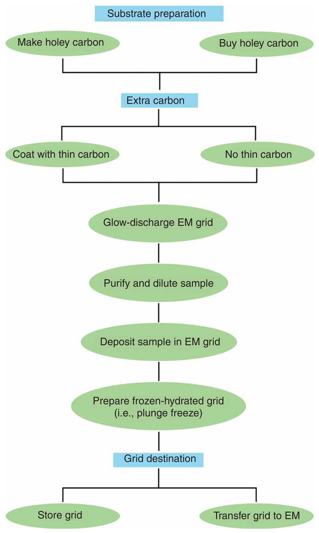
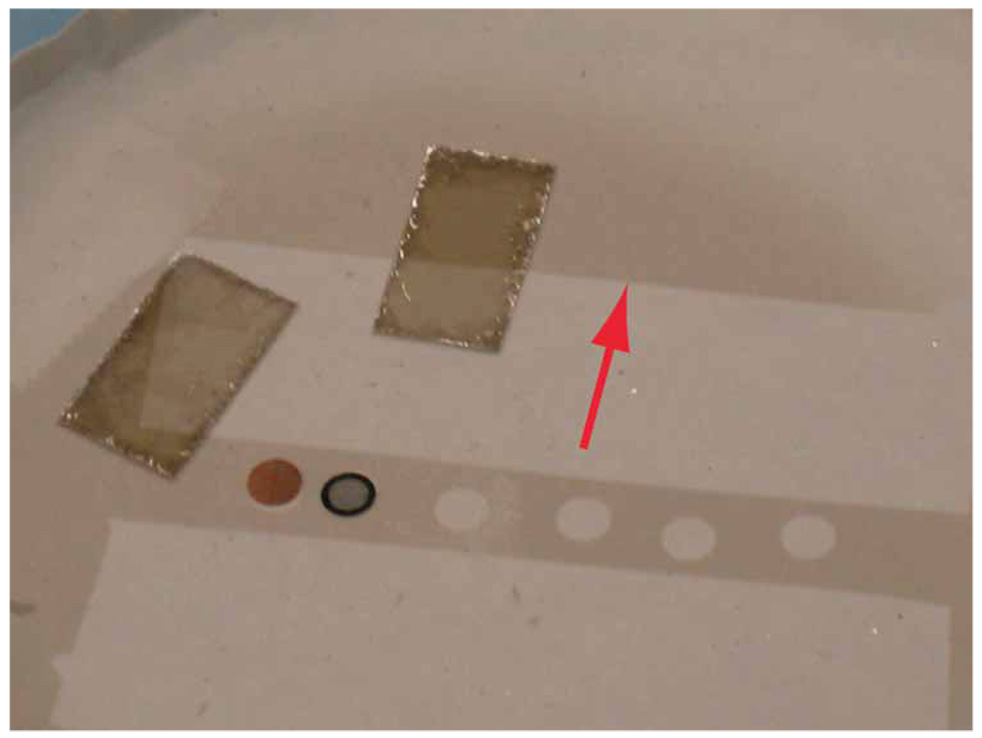
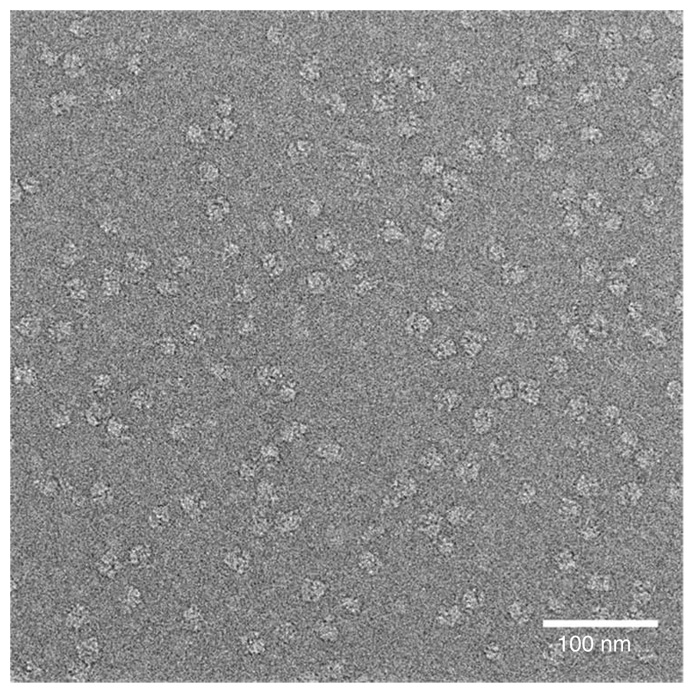
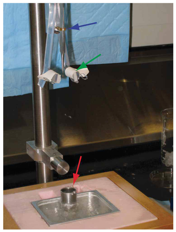
This protocol describes the preparation of frozen-hydrated single-particle specimens of macromolecular complexes. First, it describes how to create a grid surface coated with holey carbon by first inducing holes in a Formvar film to act as a template for the holey carbon that is stable under cryo-electron microscopy (cryo-EM) conditions and is sample-friendly. The protocol then describes the steps required to deposit the homogeneous sample on the grid and to plunge-freeze the grid into liquid ethane at the temperature of liquid nitrogen, so that it is suitable for cryo-EM visualization. It takes 4-5 h to make several hundred holey carbon grids and about 1 h to make the frozen-hydrated grids. The time required for sample purification varies from hours to days, depending on the sample and the specific procedure required. A companion protocol details how to collect cryo-EM data using an FEI Tecnai transmission electron microscope that can subsequently be processed to obtain a three-dimensional reconstruction of the macromolecular complex.
Figures
References
-
- Sjoberg A, Onnerfjord P, Morgelin M, Heinegard D, Blom AM. The extracellular matrix and inflammation: fibromodulin activates the classical pathway of complement by directly binding C1q. J. Biol. Chem. 2005;280:32301–32308. - PubMed
-
- Spahn CM, et al. Hepatitis C virus IRES RNA-induced changes in the conformation of the 40s ribosomal subunit. Science. 2001;291:1959–1962. - PubMed
-
- Henderson R. The potential and limitations of neutrons, electrons and X-rays for atomic resolution microscopy of unstained biological molecules. Q. Rev. Biophys. 1995;28:171–193. - PubMed
-
- Halic M, Becker T, Frank J, Spahn CM, Beckmann R. Localization and dynamic behavior of ribosomal protein L30e. Nat. Struct. Mol. Biol. 2005;12:467–468. - PubMed
-
- Jiang W, Ludtke SJ. Electron cryomicroscopy of single particles at subnanometer resolution. Curr. Opin. Struct. Biol. 2005;15:571–577. - PubMed
Publication types
MeSH terms
Substances
Grants and funding
LinkOut - more resources
Full Text Sources
Other Literature Sources
