. 2008 Jan;118(1):111-23.
doi: 10.1172/JCI29900.

TRAIL-R deficiency in mice promotes susceptibility to chronic inflammation and tumorigenesis

Affiliations

TRAIL-R deficiency in mice promotes susceptibility to chronic inflammation and tumorigenesis

Niklas Finnberg et al. J Clin Invest. 2008 Jan.

Abstract

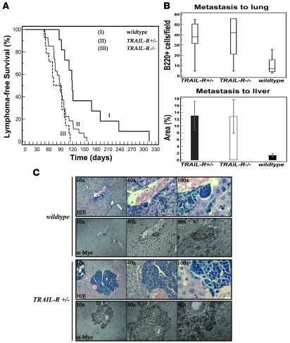
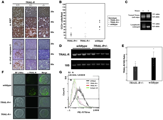
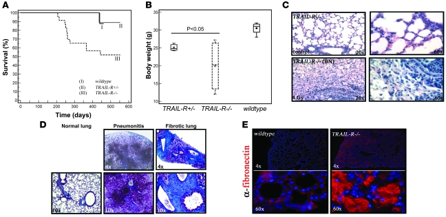
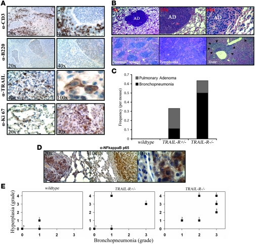
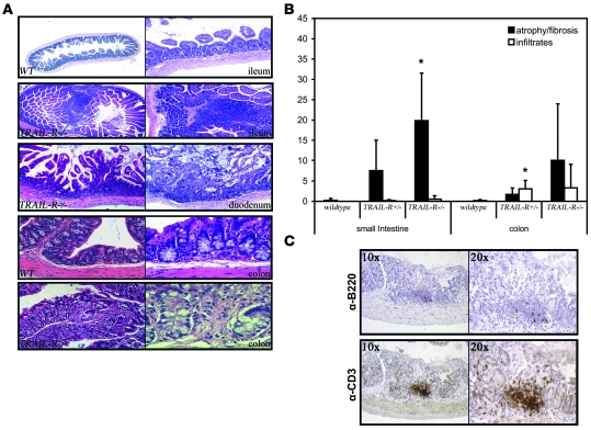
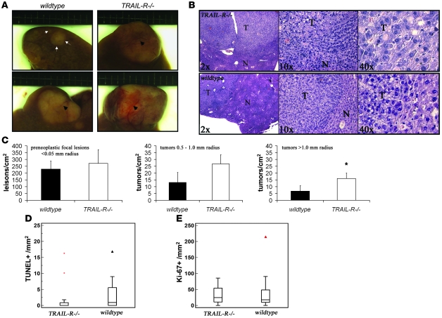
Preclinical data support the potential of the death-signaling receptors for TRAIL as targets for cancer therapy. However, it is unclear whether these death-signaling receptors suppress the emergence and growth of malignant tumors in vivo. Herein we show that TNF-related apoptosis-inducing ligand receptor (TRAIL-R), the only proapoptotic death-signaling receptor for TRAIL in the mouse, suppresses inflammation and tumorigenesis. Loss of a single TRAIL-R allele on the lymphoma-prone Emu-myc genetic background significantly reduced median lymphoma-free survival. TRAIL-R-deficient lymphomas developed with equal frequency irrespective of mono- or biallelic loss of TRAIL-R, had increased metastatic potential, and showed apoptotic defects relative to WT littermates. In addition, TRAIL-R-/- mice showed decreased long-term survival following a sublethal dose of ionizing radiation. Histological evaluation of moribund irradiated TRAIL-R-/- animals showed hallmarks of bronchopneumonia as well as tumor formation with increased NF-kappaB p65 expression. TRAIL-R also suppressed diethylnitrosamine-induced (DEN-induced) hepatocarcinogenesis, as an increased number of large tumors with apoptotic defects developed in the livers of DEN-treated TRAIL-R-/- mice. Thus TRAIL-R may function as an inflammation and tumor suppressor in multiple tissues in vivo.


Figures

Figure 1
Figure 1. Monoallelic loss of TRAIL-R promotes c-myc–driven lymphomagenesis.
(A) Kaplan-Meier survival curves for the different genotypes (TRAIL-R–/–, n = 14; TRAIL-R+/–, n = 30; and WT, n = 11) on the Eμ-myc genetic background. Survival was markedly decreased in TRAIL-R+/– and TRAIL-R–/– mice (log rank test, P = 0.023 and P = 0.003, respectively) relative to WT littermates. Immunohistochemistry for c-myc+ (α-myc) (B and C) and B220+ showed increased metastasis of lymphoma cells (B and data not shown; n = 6/genotype) to liver (n = 5/genotype; average tumor emboli area percentage ± SEM; Student’s t test, P < 0.05) and lung (box-and-whisker plot shows relative median number of B220+ cells/field; Mann-Whitney U test, P < 0.05) in both TRAIL-R+/– and TRAIL-R–/– animals relative to WT animals on the Eμ-myc genetic background.
Figure 2
Figure 2. TRAIL-R+/– lymphomas show apoptotic defects and reduced TRAIL-R mRNA expression.
(A) Immunohistochemistry shows abundant labeling of the proliferation marker Ki-67 (DAB, brown staining) in both WT (+/+) and TRAIL-R–deficient (+/– and –/–) lymphomas. Immunohistochemical staining (A) for cleaved caspase-3 and quantification thereof (B) shows fewer median number of cells expressing active caspase-3 (DAB, brown staining) in TRAIL-R+/– and TRAIL-R–/– lymphomas compared with that of WT lymphomas (Mann-Whitney U test, P < 0.05). (C) LOH was detected by PCR analysis of DNA isolated from Eμ-myc TRAIL-R+/– lymphomas in 20% (2 of 10) of the lymphomas (data not shown). (D) RT-PCR analysis on RNA isolated from Eμ-myc lymphomas of different genotypes shows decreased TRAIL-R mRNA expression in Eμ-myc TRAIL-R+/–lymphomas compared with RNA isolated from WT Eμ-myc lymphomas. (E) Relative quantitative RT-PCR analysis and densitometry shows that loss of 1 TRAIL-R allele reduced the mean (± SEM) TRAIL-R expression to approximately 60% that in WT lymphomas (n = 4 each genotype; Student’s t test, P < 0.05). (F) Immunofluorescence (FITC, green) on living Eμ-myc lymphoma cells of different TRAIL-R genotypes using the MD-5 antibody suggest expression on WT Eμ-myc lymphoma cells (α–TRAIL-R) with a heterogenous (speckled) membrane distribution as evident from the 2 different focal images. However, expression was barely detectable in TRAIL-R+/– Eμ-myc lymphoma cells and not present in TRAIL-R–/– Eμ-myc lymphoma cells. Representative images are shown from 2 independent lymphomas per genotype. Original magnification, ×100. (G) Flow cytometry analysis on Eμ-myc lymphoma cells suggests expression of TRAIL-R in WT cells but not TRAIL-R+/– and TRAIL-R–/– Eμ-myc lymphoma cells. Eμ-myc lymphoma cells from at least 2 different animals/genotype were analyzed.
Figure 3
Figure 3. WT Eμ-myc lymphomas are resistant to TRAIL in vitro and express high levels of FLIPs.
(A) Challenging isolated WT Eμ-myc lymphoma cells in vitro with TRAIL resulted in lack of cell death (as determined by tryphan blue staining) in comparison with treatment with etoposide. (B) In vitro cell death assays. Propidium iodide uptake (red staining) and fluorescent-labeled inhibitor of caspase activity (FLICA or FITC-VAD-FMK; green staining) suggest that TRAIL (1 μg/ml) only weakly triggers caspase activity and cell death relative to control in comparison with treatment with etoposide (1 μM) in living Eμ-myc lymphoma cells. (C) Total RNA isolated from WT western blot of splenic and thymic lymphomas shows increased expression of c-FLIPS in WT relative to TRAIL-R+/– lymphomas. (D) Densitometric analysis using NIH ImageJ of western blots from lymphomas (splenic and thymic) from WT (n = 10) and TRAIL-R+/– (n = 10) mice. The ratios of the FLIPs band in relation to the actin band (loading control) is shown. The red horizontal band represents median, and the red cross represents the mean. WT lymphomas have a higher FLIPs/actin ratio (P < 0.05, Student’s t test), suggesting a relatively higher FLIPs expression compared with TRAIL-R+/– lymphomas.
Figure 4
Figure 4. Expression profiling of Eμ-myc lymphomas suggests that differentially expressed genes are regulated independent of TRAIL-R gene dosage as well as a role for Stat3 in WT lymphomas.
RNA samples from TRAIL-R+/– and TRAIL-R–/– Eμ-myc lymphomas (n = 5/genotype) were hybridized to MOE430A 2.0 Affymetrix arrays. Differentially expressed genes (upregulated >2-fold) were subjected to statistical analysis. A shows the top 4 genes that showed consistent changes over the different probe sets. Hspa1b, heat-shock protein 70; Socs3, suppressor of cytokine signaling 3; Zfpm1, friend of GATA1 (also known as Fog-1); Cdkn1a, cyclin-dependent kinase inhibitor 1a (p21). *P < 0.05, Student’s t test. (B) Immunoblotting suggests that Tyr705-phosphorylated Stat3 is elevated in WT Eμ-myc lymphomas compared with TRAIL-R–deficient lymphomas. Tyr705-phosphorylated Stat3 is expressed in a subset of cells within the lymphomas, as detected by immunohistochemistry. (C) CD244 is expressed at higher levels in Eμ-myc lymphomas of TRAIL-R+/– animals (Mann-Whitney U test, P < 0.05; n = 5/genotype). Red horizontal lines indicate medians; red crosses indicate means. (D and E) TRAIL-R–deficient lymphomas (n = 3/genotype; Mann-Whitney U test) show frequent infiltration of CD244+/c-myc cells (Cy3, green; Cy2, red) compared with WT lymphomas, suggesting expression of CD244 on nonlymphoma cells. Representative pictures are shown. Original magnification, ×60 (B); ×100 (E).
Figure 5
Figure 5. TRAIL-R–/– animals show decreased survival following exposure to a single sublethal dose (4 Gy) of ionizing radiation.
(A) Survival following 4 Gy of whole-body irradiation was decreased in the TRAIL-R–/– group (n = 23) at 28–52 weeks following irradiation in comparison with the group of WT (n = 14) animals and TRAIL-R+/– (n = 13) animals (Kaplan-Meier log-rank analysis, P = 0.010). (B) However, decreased body weight was observed in both TRAIL-R+/– (n = 3) and TRAIL-R–/– (n = 4) animals relative to WT (n = 4) animals at 28 weeks following 4 Gy of ionizing irradiation (P < 0.05, Mann-Whitney U test). No weight difference was detected between genotypes in nonirradiated animals (data not shown). (C) A representative H&E staining of the lungs from lethargic TRAIL-R–/– animals irradiated with 4 Gy at 39 weeks prior to sacrifice. Extensive inflammatory emboli in the respiratory bronchioles and increased cellularity in the interstitial space was observed (C; ×20, lower left and right panels). Lungs were histochemically stained with Masson’s trichrome (D) in order to detect the presence of collagen (bright blue) and were analyzed by immunofluorescence for fibronectin (E; Cy3, red). Lungs from irradiated and lethargic TRAIL-R–/– animals showed severe pneumonitis (D) and extensive deposition of collagen and fibronectin (E).
Figure 6
Figure 6. Loss of TRAIL-R leads to increased infiltration of CD3+ cells and tumorigenesis following sublethal irradiation.
(A) Immunohistochemistry on lungs from TRAIL-R–/– animals with infiltrates show increased numbers of CD3+ cells but little to no positive staining for B220. Infiltrates contained a number of TRAIL-positive cells expressing cytoplasmic and membrane-bound TRAIL and focal areas that stained positive for Ki-67. (B) Mice lacking 1 or 2 alleles of TRAIL-R show an increased incidence of pulmonary adenomas (top row). Microphotographs show a normal spleen and a splenic lymphoma with metastasis to the liver (bottom row). Lungs from 12 WT, 9 TRAIL-R+/–, and 18 TRAIL-R–/– mice were examined and stained immunohistochemically. (C) The frequency per mouse of pulmonary adenomas in relation to bronchopneumonia is shown. WT, n = 12; TRAIL-R+/–, n = 9; TRAIL-R–/–, n = 18. (D) Irradiated TRAIL-R–/– animals show correlation between bronchopneumonia and hyperplasia in their lungs. Hyperplastic adenomatous focal lesions (left panel) and pulmonary adenomas (middle and right panel) in the lungs of irradiated TRAIL-R–/– animals showed positive immunohistochemistry for NF-κB p65. Staining was observed in the cytoplasm and nucleus (right panel) of adenomatous cells. Representative photographs of investigated animals are shown. (E) Lungs of irradiated animals of the different TRAIL-R genotypes (WT, n = 7; TRAIL-R+/–, n = 5; TRAIL-R–/–, n = 10) were blindly classified according to inflammatory grade (bronchopneumonia grade) by the use of histological examination and immunohistochemistry for CD3 and fibronectin. Preneoplasia/neoplasia (hyperplastic) grading was based on combined macroscopic observations, histological findings (H&E staining), and immunohistochemistry for Ki-67. Grade 0, no or only scattered stained cells constituting less than 2% of the section; grade 1, heterogeneous staining with at least 20% of the section showing 2%–10% positive cells; grade 2, at least 20% of the section showing 11%–50% positive cells; grade 3, and at least 20% of the section showing more than 50% positive cells. For hyperplasia/neoplasia, presence of adenoma was classified as grade 4. The n-value represented by each data point is shown.
Figure 7
Figure 7. TRAIL-R suppresses chronic colitis following sublethal irradiation.
(A) The small intestine (ileum) of WT and TRAIL-R–/– animals sublethally irradiated 32 weeks prior to sacrifice show increased lymphoid infiltration and luminal protrusions in the TRAIL-R–/– animals compared with WT mice. The proximal part of the small bowel (duodenum) showed epithelial atrophy and erosion in an irradiated TRAIL-R–/– animal at 32 weeks following irradiation. Normal colon of a WT animal at 32 weeks following 4 Gy of irradiation is compared with TRAIL-R–/– animals, which showed chronic enterocolitis following the same treatment. Original magnification, ×4 (first and second row, first column); ×10 (first and second row, second column); ×20 (third row); ×40 (fourth row) (fifth row, first column); ×60 (fifth row, second column). (B) The surface area covered with either lymphoid infiltrates or atrophic lesions was assessed from tissue sections of the small intestine and colon from WT (n = 4), TRAIL-R+/– (n = 3), and TRAIL-R–/– (n = 4) mice subjected to 4 Gy of whole-body irradiation. *P < 0.05, Student’s t test. (C) Representative immunohistochemistry for B220 and CD3 on a TRAIL-R–/– small intestine with atrophy shows abundant infiltration of CD3+ cells.
Figure 8
Figure 8. Loss of TRAIL-R renders mice susceptible to DEN-induced hepatocarcinogenesis.
(A and B) TRAIL-R–/– mice (n = 10) injected with DEN (0.30 mmol/kg body weight) at 7 days of age showed an increased liver tumor load compared with WT littermates (n = 10) at 10 months after treatment. (A) Gross observations of small (top row, demarcated with arrows and arrowheads) and larger liver tumors in DEN-treated animals (bottom row, demarcated with arrowheads). (B) Microscopy of H&E-stained histological sections shows the location of a liver tumor (T) with surrounding normal (N) liver tissue. (C) Detailed histological analysis of livers (>0.5 cm2 liver area analyzed, excluding macroscopic lesions) from randomly selected littermates (n = 3/genotype) shows an increased number of lesions exceeding a radius of 1.0 mm in TRAIL-R–/– animals. *P < 0.05, Student’s t test. Means ± SD are shown. (D and E) Immunohistochemical analysis of proliferation (Ki-67 labeling; E) and cell death (TUNEL staining; D) in HCCs from WT (n = 17) and TRAIL-R–/– (n = 17) animals treated with DEN show reduced TUNEL labeling in TRAIL-R–/– HCCs (Mann-Whitney U test, P < 0.05) compared with WT HCCs, whereas similar levels of Ki-67–positive cells were seen in HCCs of both genotypes.

Comment in

Similar articles

Cited by

References

    1. Boldin M.P., et al. A novel protein that interacts with the death domain of Fas/APO1 contains a sequence motif related to the death domain. J. Biol. Chem. 1995;270:7795–7798. - PubMed
    1. Chinnaiyan A.M., O’Rourke K., Tewari M., Dixit V.M. FADD, a novel death domain-containing protein, interacts with the death domain of Fas and initiates apoptosis. Cell. 1995;81:505–512. - PubMed
    1. Bodmer J.L., et al. TRAIL receptor-2 signals apoptosis through FADD and caspase-8. Nat. Cell Biol. 2000;2:241–243. - PubMed
    1. Kischkel F.C., et al. Apo2L/TRAIL-dependent recruitment of endogenous FADD and caspase-8 to death receptors 4 and 5. Immunity. 2000;12:611–620. - PubMed
    1. Sprick M.R., et al. FADD/MORT1 and caspase-8 are recruited to TRAIL receptors 1 and 2 and are essential for apoptosis mediated by TRAIL receptor 2. Immunity. 2000;12:599–609. - PubMed

Publication types

MeSH terms

Substances