. 2008 Jan;118(1):124-32.
doi: 10.1172/JCI33255.

The MEF2D transcription factor mediates stress-dependent cardiac remodeling in mice

Affiliations

The MEF2D transcription factor mediates stress-dependent cardiac remodeling in mice

Yuri Kim et al. J Clin Invest. 2008 Jan.

Abstract

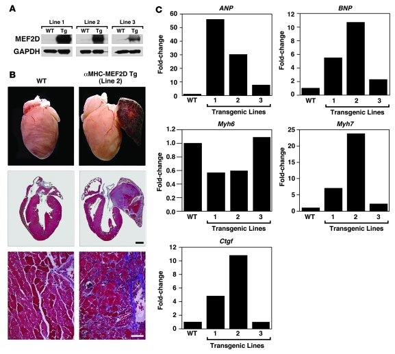
The adult heart responds to excessive neurohumoral signaling and workload by a pathological growth response characterized by hypertrophy of cardiomyocytes and activation of a fetal program of cardiac gene expression. These responses culminate in diminished pump function, ventricular dilatation, wall thinning, and fibrosis, and can result in sudden death. Myocyte enhancer factor-2 (MEF2) transcription factors serve as targets of the signaling pathways that drive pathological cardiac remodeling, but the requirement for MEF2 factors in the progression of heart disease in vivo has not been determined. MEF2A and MEF2D are the primary MEF2 factors expressed in the adult heart. To specifically determine the role of MEF2D in pathological cardiac remodeling, we generated mice with a conditional MEF2D allele. MEF2D-null mice were viable, but were resistant to cardiac hypertrophy, fetal gene activation, and fibrosis in response to pressure overload and beta-chronic adrenergic stimulation. Furthermore, we show in a transgenic mouse model that forced overexpression of MEF2D was sufficient to drive the fetal gene program and pathological remodeling of the heart. These results reveal a unique and important function for MEF2D in stress-dependent cardiac growth and reprogramming of gene expression in the adult heart.


Figures

Figure 1
Figure 1. Generation of mice with a conditional Mef2d mutation.
(A) Schematic representation of the mouse Mef2d locus and targeting strategy. Positions of 3ι and 5ι probes used for Southern blots are shown. The positions of the PCR primers used for genotyping mutant alleles are marked with arrows (circles labeled 1, 2, and 3). C, ClaI; frt, FLP recombinase target; E, EcoRI; TAD, transactivation domain; MADS, MCM1, agamous, deficiens, serum response factor; N, NcoI; X, XhoI. (B) Southern blot analysis of Mef2d mutant alleles. Genomic DNA was digested with EcoRI and hybridized to a 5ι probe in the left panel and to a 3ι probe in the right panel. WT, wild-type allele; Mef2dneo-loxP, conditional allele; Mef2dloxP, conditional allele with the Neo cassette removed; Mef2dKO, null allele. (C) PCR genotyping to distinguish different Mef2d alleles. The positions of the primers that produce these PCR products are labeled (1, 2, and 3) and circled on A. All 3 primers were added to the PCR reactions and the PCR products were loaded into lanes 2–7. (D) Expression of wild-type and mutant Mef2d detected by RT-PCR. Mef2d mutant allele lacks exon 3, which encodes the MADS- and MEF2-specific domains. GAPDH was used as a loading control. Labels on the left side of the panel indicate exon location and direction of primers used for RT-PCR. (E) Western blot analysis to detect WT and mutant MEF2D (KO) proteins. The mutant MEF2D protein is truncated due to deletion of exon 3. α-Actin protein was used as a loading control. (F) Expression level of Mef2 detected by quantitative PCR. Error bars indicate ±SEM. u.d., undetectable.
Figure 2
Figure 2. Blunted hypertrophy of Mef2d mutant mice following TAC.
(A) HW/TL ratios (±SEM) of WT and Mef2d mutant mice were determined 21 days after TAC. (B) Hearts from WT and Mef2d mutant mice subjected to either a sham operation or pressure overload (TAC) are shown at the top. Histological sections stained with Masson’s trichrome are shown on the bottom. Masson’s trichrome staining of WT and Mef2d mutant hearts indicates lack of fibrosis in Mef2d–/– hearts in response to pressure overload by TAC. Scale bars: 1 mm (middle panel); 40 μm (bottom panel). (C) Mean cross-sectional area of cardiomyocytes (±SEM) in WT and Mef2d mutant mice was measured 21 days after TAC. (D) Using morphometric analysis of picrosirius red stained heart sections, the amount of myocardial fibrosis was assessed. Fibrosis was apparent in the hearts of WT mice following TAC. In contrast, virtually no fibrosis was detected in the hearts of Mef2d mutant mice. Values indicate fold changes of fibrosis in each group compared with a group of sham-operated WT mice (±SEM).
Figure 3
Figure 3. Echocardiographic analysis of Mef2d mutant mice following TAC.
Mef2d mutant mice display less LV dilation during diastole and a less pronounced decrease in FS in response to TAC than WT mice.
Figure 4
Figure 4. Resistance of Mef2d mutant mice to fetal gene activation following TAC.
Expression of cardiac hypertrophic markers, fibrosis markers, Mef2a, and Mef2d in WT and Mef2d mutant hearts was detected by quantitative PCR 21 days after TAC (n = 3–5 per group). Values indicate expression level relative to a WT sham-operated group (±SEM). ANP, atrial natriuretic peptide; BNP, brain natriuretic peptide; Myh6, myosin, heavy polypeptide 6, cardiac muscle, alpha; Myh7, myosin, heavy polypeptide 7, cardiac muscle, beta; Acta1, actin, alpha 1, skeletal muscle; Ctgf, connective tissue growth factor; Col1a2, procollagen, type I, alpha 2; Col3a1, procollagen, type III, alpha 1; n.d., non-determined.
Figure 5
Figure 5. Decreased hypertrophic response of Mef2d mutant mice following β-adrenergic stimulation.
(A) WT and Mef2d mutant mice were chronically infused with either saline vehicle or ISO (8.8 mg/kg/day) for 7 days and sacrificed for assessment of cardiac remodeling. HW/TL ratios (±SEM) are shown as bar graphs (n = 7–9 per group). n.s., non-significant. (B) Whole hearts of WT and Mef2d mutant mice treated with either saline vehicle or ISO (8.8 mg/kg/day) are shown at the top. Hearts were sectioned and stained with H&E as seen on bottom. Scale bar: 1 mm. (C) Representative bar graphs indicating less LV dilation and a decrease in FS in Mef2d mutant mice compared with WT mice in response to ISO treatment.
Figure 6
Figure 6. Pathological cardiac remodeling induced by overexpression of MEF2D.
(A) Proteins were extracted from ventricles of α-MHC-MEF2D transgenic mouse lines at 6 weeks to 8 weeks of age for Western blot analysis using an antibody against MEF2D. GAPDH was used as a loading control. (B) Hearts were dissected from α-MHC-MEF2D male mice (line 2) and WT littermates at 8 weeks of age. Note the enlarged left atrium in the α-MHC-MEF2D heart. The lower panels are histological sections stained with Masson’s trichrome to detect fibrosis. Scale bar: 1 mm (middle panel); 40 μm (bottom panel). (C) Expression level of various hypertrophy and fibrosis markers was evaluated by quantitative PCR in each transgenic line. Note the correlation between expression level of MEF2D (A) and that of hypertrophy and fibrosis markers.

Similar articles

Cited by

References

    1. Czubryt M.P., Olson E.N. Balancing contractility and energy production: the role of myocyte enhancer factor 2 (MEF2) in cardiac hypertrophy. Recent Prog. Horm. Res. 2004;59:105–124. - PubMed
    1. Frey N., Katus H.A., Olson E.N., Hill J.A. Hypertrophy of the heart: a new therapeutic target? Circulation. 2004;109:1580–1589. - PubMed
    1. Frey N., Olson E.N. Cardiac hypertrophy: the good, the bad, and the ugly. Annu. Rev. Physiol. 2003;65:45–79. - PubMed
    1. McKinsey T.A., Olson E.N. Toward transcriptional therapies for the failing heart: chemical screens to modulate genes. J. Clin. Invest. 2005;115:538–546. - PMC - PubMed
    1. Backs J., Olson E.N. Control of cardiac growth by histone acetylation/deacetylation. Circ. Res. 2006;98:15–24. - PubMed

Publication types

MeSH terms