

Cholinergic interneurons are differentially distributed in the human striatum
- PMID: 18080007
- PMCID: PMC2137841
- DOI: 10.1371/journal.pone.0001174
Cholinergic interneurons are differentially distributed in the human striatum
Abstract
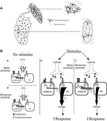
Background: The striatum (caudate nucleus, CN, and putamen, Put) is a group of subcortical nuclei involved in planning and executing voluntary movements as well as in cognitive processes. Its neuronal composition includes projection neurons, which connect the striatum with other structures, and interneurons, whose main roles are maintaining the striatal organization and the regulation of the projection neurons. The unique electrophysiological and functional properties of the cholinergic interneurons give them a crucial modulating function on the overall striatal response.
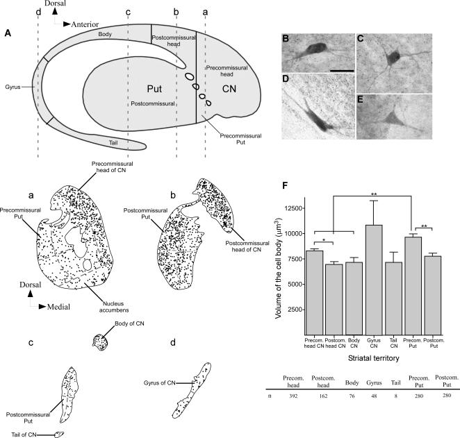
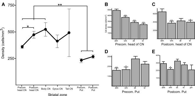
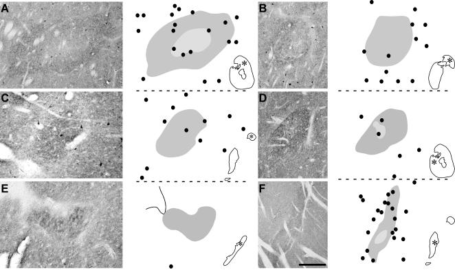
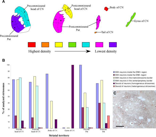
Methodology/principle findings: This study was carried out using stereological methods to examine the volume and density (cells/mm(3)) of these interneurons, as visualized by choline acetyltransferase (ChAT) immunoreactivity, in the following territories of the CN and Put of nine normal human brains: 1) precommissural head; 2) postcommissural head; 3) body; 4) gyrus and 5) tail of the CN; 6) precommissural and 7) postcommissural Put. The distribution of ChAT interneurons was analyzed with respect to the topographical, functional and chemical territories of the dorsal striatum. The CN was more densely populated by cholinergic neurons than the Put, and their density increased along the anteroposterior axis of the striatum with the CN body having the highest neuronal density. The associative territory of the dorsal striatum was by far the most densely populated. The striosomes of the CN precommissural head and the postcommissural Put contained the greatest number of ChAT-ir interneurons. The intrastriosomal ChAT-ir neurons were abundant on the periphery of the striosomes throughout the striatum.
Conclusions/significance: All these data reveal that cholinergic interneurons are differentially distributed in the distinct topographical and functional territories of the human dorsal striatum, as well as in its chemical compartments. This heterogeneity may indicate that the posterior aspects of the CN require a special integration of information by interneurons. Interestingly, these striatal regions have been very much left out in functional studies.
Conflict of interest statement
Figures

Similar articles
-
Distribution of GABAergic interneurons and dopaminergic cells in the functional territories of the human striatum.PLoS One. 2012;7(1):e30504. doi: 10.1371/journal.pone.0030504. Epub 2012 Jan 17. PLoS One. 2012. PMID: 22272358 Free PMC article.
-
Morphological features, distribution and compartmental organization of the nicotinamide adenine dinucleotide phosphate reduced-diaphorase interneurons in the human striatum.J Comp Neurol. 2005 Aug 29;489(3):311-27. doi: 10.1002/cne.20616. J Comp Neurol. 2005. PMID: 16025450
-
Neuronal density and proportion of interneurons in the associative, sensorimotor and limbic human striatum.Brain Struct Funct. 2018 May;223(4):1615-1625. doi: 10.1007/s00429-017-1579-8. Epub 2017 Nov 28. Brain Struct Funct. 2018. PMID: 29185108
-
Chemical anatomy of striatal interneurons in normal individuals and in patients with Huntington's disease.Brain Res Brain Res Rev. 2000 Nov;34(1-2):80-101. doi: 10.1016/s0165-0173(00)00039-4. Brain Res Brain Res Rev. 2000. PMID: 11086188 Review.
-
Cholinergic interneuron characteristics and nicotinic properties in the striatum.J Neurobiol. 2002 Dec;53(4):590-605. doi: 10.1002/neu.10150. J Neurobiol. 2002. PMID: 12436423 Review.
Cited by
-
Compartmental function and modulation of the striatum.J Neurosci Res. 2019 Dec;97(12):1503-1514. doi: 10.1002/jnr.24522. Epub 2019 Sep 5. J Neurosci Res. 2019. PMID: 31489687 Free PMC article. Review.
-
Mapping Dorsal and Ventral Caudate in Older Adults: Method and Validation.Front Aging Neurosci. 2017 Apr 4;9:91. doi: 10.3389/fnagi.2017.00091. eCollection 2017. Front Aging Neurosci. 2017. PMID: 28420985 Free PMC article.
-
Distribution of GABAergic interneurons and dopaminergic cells in the functional territories of the human striatum.PLoS One. 2012;7(1):e30504. doi: 10.1371/journal.pone.0030504. Epub 2012 Jan 17. PLoS One. 2012. PMID: 22272358 Free PMC article.
-
Differential striatal spine pathology in Parkinson's disease and cocaine addiction: a key role of dopamine?Neuroscience. 2013 Oct 22;251:2-20. doi: 10.1016/j.neuroscience.2013.07.011. Epub 2013 Jul 16. Neuroscience. 2013. PMID: 23867772 Free PMC article. Review.
-
α-Synuclein pathology accumulates in sacral spinal visceral sensory pathways.Ann Neurol. 2015 Jul;78(1):142-9. doi: 10.1002/ana.24430. Epub 2015 May 25. Ann Neurol. 2015. PMID: 25893830 Free PMC article.
References
-
- Parent A. Extrinsic connections of the basal ganglia. Trends Neurosci. 1990;13:254–258. - PubMed
-
- Haber SN. The primate basal ganglia: parallel and integrative networks. J Chem Neuroanat. 2003;26:317–330. - PubMed
-
- DiFiglia M, Pasik P, Pasik T. A Golgi study of neuronal types in the neostriatum of monkeys. Brain Res. 1976;114:245–256. - PubMed
-
- Yelnik J, Francois C, Percheron G, Tande D. Morphological taxonomy of the neurons of the primate striatum. J Comp Neurol. 1991;313:273–294. - PubMed
Publication types
MeSH terms
Substances
LinkOut - more resources
Full Text Sources
