. 2008 May;103(3):244-56.
doi: 10.1007/s00395-007-0693-9. Epub 2007 Dec 13.

Regulation of cell-matrix contacts and beta-catenin signaling in VSMC by integrin-linked kinase: implications for intimal thickening

Affiliations

Regulation of cell-matrix contacts and beta-catenin signaling in VSMC by integrin-linked kinase: implications for intimal thickening

Amrita Dwivedi et al. Basic Res Cardiol. 2008 May.

Abstract

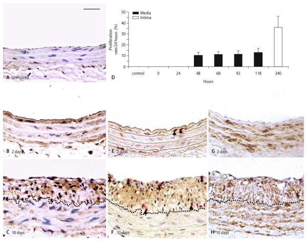
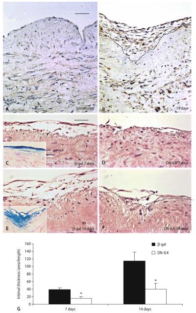
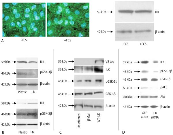
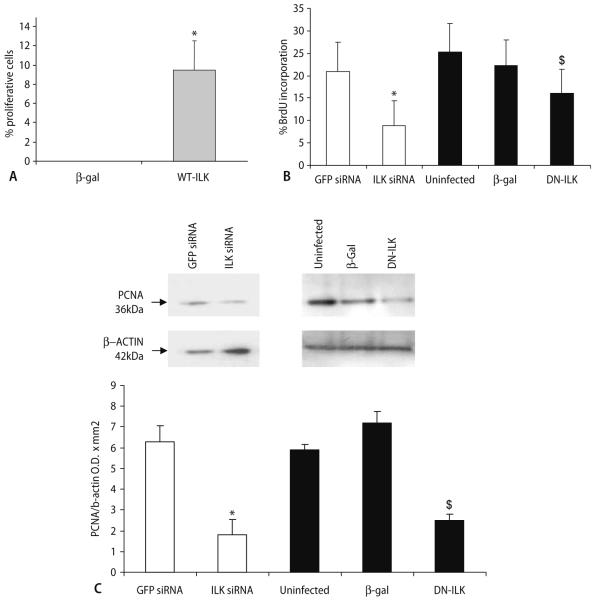
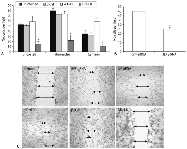
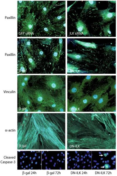
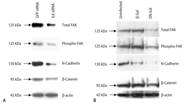
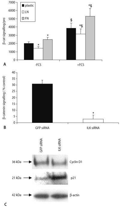
Vascular smooth muscle cell (VSMC) proliferation and migration is responsible for intimal thickening that occurs in restenosis and atherosclerosis. Integrin-linked kinase (ILK) is a serine/threonine protein kinase implicated in signaling pathways involved in cell proliferation and migration. We studied the involvement of ILK in intimal thickening. ILK expression was significantly increased in two models of intimal thickening: balloon-injured rat carotid arteries and human saphenous vein organ cultures. Over-expression of a dominant negative ILK (DN-ILK) significantly reduced intimal thickening by approximately 50% in human saphenous vein organ cultures, demonstrating an important role in intimal thickening. ILK protein and activity was reduced on laminin and up-regulated on fibronectin, indicating ILK protein expression is modulated by extracellular matrix composition. Inhibition of ILK by siRNA knockdown and DN-ILK significantly decreased VSMC proliferation and migration while wild type ILK significantly increased proliferation and migration on laminin, confirming an essential role of ILK in both processes. Localization of paxillin and vinculin and protein levels of FAK and phospho-FAK indicated that inhibition of ILK reduced focal adhesion formation. Additionally, inhibition of ILK significantly attenuated the presence of the cell-cell complex proteins N-cadherin and beta-catenin, and beta-catenin signaling. We therefore suggest ILK modulates VSMC proliferation and migration at least in part by acting as a molecular scaffold in focal adhesions as well as modulating the stability of cell-cell contact proteins and beta-catenin signaling. In summary, ILK plays an important role in intimal thickening by modulating VSMC proliferation and migration via regulation of cell-matrix and cell-cell contacts and beta-catenin signaling.


Figures

Fig. 1
Fig. 1
Integrin-linked kinase (ILK) is elevated during intimal thickening. Representative immunohistochemical staining for ILK (brown) in rat carotid arteries from sham-operated controls (A), 2 days (B), and 10 days (C) after balloon injury. Quantification of proliferation in media (black bars) and intima (white bar) at various time points after injury assessed by BrdU incorporation 24 h prior to carotid artery removal (D). Dual immunohistochemistry for ILK (brown) and BrdU (red) at 2 days (E) and 10 days (F). Immunohistochemistry for smooth muscle α-actin (brown) at 2 days (G), and 10 days (H). Dotted lines indicate the intima:media boundary. Nuclei are stained blue. Scale bar in A represents 50 μm and applies to all panels. n = 4 per time-point
Fig. 2
Fig. 2
Integrin-linked kinase modulates intimal thickening. Representative (n = 4) immunohistochemical staining for ILK (brown) of uncultured saphenous vein (A) and saphenous vein after 14 days of culture (B). Scale bar in A represents 100 μm and applies to A and B. Dotted lines indicate the intima:media boundary. Representative sections from human saphenous vein infected with RAdLacZ (β-gal: C, E) and RAdDN-ILK (D, F), stained with haematoxylin and eosin, 7 (C, D) and 14 (E, F) days after infection. Dotted lines indicate intima:media boundary. Scale bar in C represents 50 μm and applies to C–F. Inset in C and E show X-gal staining of saphenous vein; blue colour identifies β-galactosidase expression. G Quantification of intimal thickness in RAdDN-ILK infected segments, asterisk indicates significant difference from β-gal control (P < 0.05, n = 5)
Fig. 3
Fig. 3
Modulation of ILK in cultured VSMCs. Immunocytochemistry and Western blotting for ILK in VSMCs cultured in presence (+FCS) or absence (−)FCS) of serum (A). Scale bar represents 10 μm; green colour indicates ILK and nuclei are stained blue with DAPI. Representative Western blots for ILK and phosphorylated GSK-3β in lysates of VSMCs cultured in serum-free media for 24 h on tissue culture plastic or laminin, n = 3 (B). Western blotting for V5-tag, ILK, phosphorylated GSK-3β and total GSK-3β in lysates of uninfected VSMCs and VSMCs infected with RAdLacZ (β-gal) and RAdWT-ILK (C). Representative Western blots for ILK, phosphorylated GSK-3β, total GSK-3β, phosphorylated Akt, and total Akt in lysates of VSMCs treated with GFP siRNA and ILK siRNA (n = 5) (D). β-actin is shown as a loading control.
Fig. 4
Fig. 4
Integrin-linked kinase modulates proliferation. Proliferation assessed by BrdU incorporation in VSMCs infected with RAdLacZ (β-gal) or RAdWT-ILK and then cultured for 24 h on laminin in serum-free media (A). Asterisk indicates significant difference from β-gal control, P < 0.05, n = 3. Proliferation assessed by BrdU incorporation (B) and representative Western blotting for PCNA and β-actin (C) in VSMCs treated with GFP siRNA and ILK siRNA and uninfected VSMCs, and VSMCs infected with RAd-LacZ (β-gal) and RAd DN-ILK and then cultured for 24 h in FCS. The number of BrdU positive cells is expressed as a percentage of the total. Asterisk indicates significant difference from GFP siRNA and dollar indicates significant difference from uninfected and β-gal controls, P < 0.05, n = 3 and n = 4, respectively
Fig. 5
Fig. 5
Integrin-linked kinase modulates migration. Migration of VSMCs infected with RAdLacZ (β-gal) RAd DN-ILK or RAdWT-ILK and then placed in uncoated, or fibronectin- or laminin-coated chemotaxis chambers for 24 h (A). Asterisk indicates significant difference from β-gal control and uninfected control, P < 0.05, n = 3. Migration assessed by 24 h in uncoated chemotaxis chambers in VSMCs treated with GFP siRNA and ILK siRNA (B). Migration assessed 18 h after wounding in VSMCs treated with GFP siRNA and ILK siRNA and uninfected VSMCs, and VSMCs infected with RAdLacZ (β-gal) and RAd DN-ILK (C). Asterisk indicates significant difference from GFP siRNA control, P < 0.05, n = 3. Scale bar represents 250 μm
Fig. 6
Fig. 6
Effect of DN-ILK on focal adhesion proteins and cell viability. Immunocytochemical staining for paxillin, vinculin, and α-actin in VSMCs treated with GFP siRNA and ILK siRNA, and VSMCs infected with RAdLacZ (β-gal) and RAdDN-ILK (DN-ILK) 24 h after the infection period. Immunocyto-chemical staining for cleaved caspase-3 in VSMCs infected with RAdLacZ and RAdDN-ILK 24 and 72 h after the infection period. Arrows indicate positive cells. Green colour indicates the presence of protein; nuclei are stained blue with DAPI in all panels except α-actin. Scale bars represent 20 μm
Fig. 7
Fig. 7
Dominant negative-ILK reduces focal adhesions and cell–cell contacts. Western blots for total FAK, phosphorylated FAK, N-cadherin, β-catenin, and β-actin at 24 h after treatment with GFP siRNA and ILK siRNA (A) or 24 h after infection period in uninfected VSMCs and VSMCs infected with RAd LacZ (β-gal) and RAd DN-ILK (B). Numbers represent molecular weights of proteins, n = 8 DN-ILK and n = 5 siRNA
Fig. 8
Fig. 8
Integrin-linked kinase regulates β-catenin signaling. β-Catenin signaling in VSMCs grown on plastic, laminin or fibronectin in the presence (+FCS) or absence of FCS (−FCS) for 24 h. Asterisk and dollar indicate significant difference (P < 0.05) from plastic and −FCS, respectively, n = 3. Induction of β-catenin signaling 24 h after the addition of FCS was assessed in TOPGAL VSMCs subjected to GFP or ILK siRNA. Results are expressed as a percentage of the quiescent VSMCs (control), n = 3, asterisk indicates significant difference from GFP siRNA P < 0.05 (B). Western blots for cyclin D1, p21 and β-actin at 24 h after treatment with GFP siRNA and ILK siRNA. Numbers represent molecular weights of proteins, n = 3 (C)

Similar articles

Cited by

References

    1. Bar H, Wende P, Watson L, Denger S, Van Eys G, Kreuzer J, Jahn L. Smoothelin is an indicator of reversible phenotype modulation of smooth muscle cells in balloon-injured rat carotid arteries. Basic Res Cardiol. 2002;97:9–16. - PubMed
    1. Bou-Gharios G, Ponticos M, Rajkumar V, Abraham D. Extra-cellular matrix in vascular networks. Cell Prolif. 2004;37:207–220. - PMC - PubMed
    1. Chen X, Gumbiner BM. Cross-talk between different adhesion molecules. Curr Opin Cell Biol. 2006;18:572–578. - PubMed
    1. Delcommenne M, Tan C, Gray V, Rue L, Woodgett J, Dedhar S. Phos-phoinositide-3-OH kinase-dependent regulation of glycogen synthase kinase 3 and protein kinase B/AKT by the integrin-linked kinase. Proc Natl Acad Sci USA. 1998;95:11211–11216. - PMC - PubMed
    1. Dietrich T, Perlitz C, Licha K, Stawowy P, Atrott K, Tachezy M, Meyborg H, Stocker C, Grafe M, Fleck E, Schirner M, Graf K. ED-B fibronectin (ED-B) can be targeted using a novel single chain antibody conjugate and is associated with macrophage accumulation in atherosclerotic lesions. Basic Res Cardiol. 2007;102:298–307. - PubMed

Publication types

MeSH terms