

PEComas: the past, the present and the future
- PMID: 18080139
- PMCID: PMC2234444
- DOI: 10.1007/s00428-007-0509-1
PEComas: the past, the present and the future
Abstract
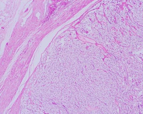
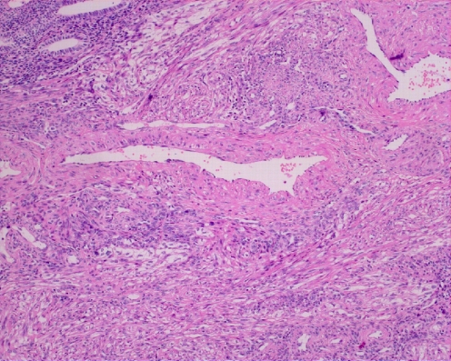
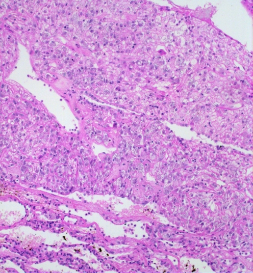
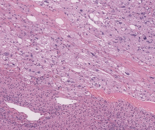
The perivascular epithelioid cell (PEC) is a cell type constantly present in a group of tumors called PEComas. PEC expresses myogenic and melanocytic markers, such as HMB45 and actin. Recently, recurrent chromosomal alterations have been demonstrated in PEC. At present, PEComa is a widely accepted entity. In the past 10 years, the use of this term has allowed to report and describe numerous cases permitting to start highlighting the biology of this group of lesions. PEComas are related to the genetic alterations of tuberous sclerosis complex (TSC), an autosomal dominant genetic disease due to losses of TSC1 (9q34) or TSC2 (16p13.3) genes which seem to have a role in the regulation of the Rheb/mTOR/p70S6K pathway. There are some open questions about PEComas regarding its histogenesis, the definition of epithelioid angiomyolipoma and the identification of the histological criteria of malignancy. An innovative therapeutic trial using rapamycin is under way for tumors occurring in TSC such as renal angiomyolipoma and lymphangioleiomyomatosis. Its success could provide the rationale for the use of the same drug in other lesions composed of PECs, especially in the malignant ones.
Figures


References
-
- Abdulla M, Bui HX, del Rosario AD, Ross JS (1994) Renal angiomyolipoma. DNA content and immunohistochemical study of classic and multicentric variants. Arch Pathol Lab Med 118:735–739 - PubMed
-
- Agaimy A, Wünsch PH (2006) Perivascular epithelioid cell sarcoma (malignant PEComa) of the ileum. Pathol Res Pract 202:37–41 - PubMed
-
- Al-Saleem T, Wessner LL, Scheithauer BW, Patterson K, Roach ES, Dreyer SJ, Fujikawa K, Bjornsson J, Bernstein J, Henske EP (1998) Malignant tumors of the kidney, brain, and soft tissues in children and young adults with the tuberous sclerosis complex. Cancer 83:2208–2216 - PubMed
-
- Anderson AE, Yang X, Young RH (2002) Epithelioid angiomyolipoma of the ovary: a case report and literature review. Int J Gynecol Pathol 21:69–73 - PubMed
-
- Apitz K (1943) Die Geschwülste und Gewebsmissbildungen der Nierenrinde. II Midteilung. Die mesenchymalen Neubildungen. Virchows Arch 311:306–327
Publication types
MeSH terms
Substances
LinkOut - more resources
Full Text Sources
Other Literature Sources
Medical
Miscellaneous
