. 2008 Jan;38(1):20-9.
doi: 10.1002/eji.200737799.

Broad T cell immunity to the LcrV virulence protein is induced by targeted delivery to DEC-205/CD205-positive mouse dendritic cells

Affiliations

Broad T cell immunity to the LcrV virulence protein is induced by targeted delivery to DEC-205/CD205-positive mouse dendritic cells

Yoonkyung Do et al. Eur J Immunol. 2008 Jan.

Abstract

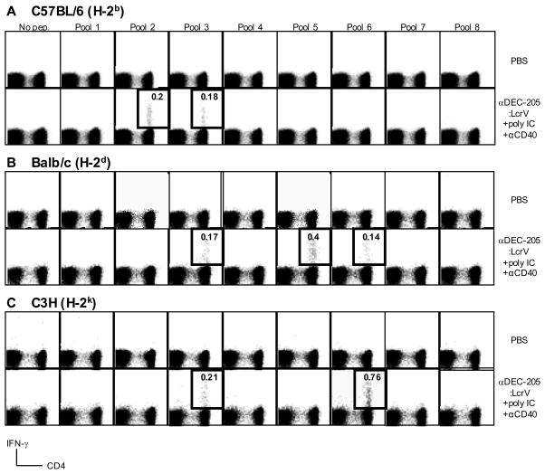
There is a need for a more efficient vaccine against the bacterium Yersinia pestis, the agent of pneumonic plague. The F1-LcrV (F1-V) subunit vaccine in alhydrogel is known to induce humoral immunity. In this study, we utilized DC to investigate cellular immunity. We genetically engineered the LcrV virulence protein into the anti-DEC-205/CD205 mAb and thereby targeted the conjugated protein directly to mouse DEC-205(+) DC in situ. We observed antigen-specific CD4(+) T cell immunity measured by intracellular staining for IFN-gamma in three different mouse strains (C57BL/6, BALB/c, and C3H/HeJ), while we could not observe such T cell responses with F1-V vaccine in alhydrogel. Using a peptide library for LcrV protein, we identified two or more distinct CD4(+) T cell mimetopes in each MHC haplotype, consistent with the induction of broad immunity. When compared to nontargeted standard protein vaccine, DC targeting greatly increased the efficiency for inducing IFN-gamma-producing T cells. The targeted LcrV protein induced antibody responses to a similar extent as the F1-V subunit vaccine, but Th1-dependent IgG2a and IgG2c isotypes were observed only after anti-DEC-205:LcrV mAb immunization. This study sets the stage for the analysis of functional roles of IFN-gamma-producing T cells in Y. pestis infection.


Conflict of interest statement

CONFLICT OF INTEREST STATEMENT

R. M. Steinman is a consultant to Celldex, which is developing human DEC-205-based vaccines. Other authors have no conflict of interest.

Figures

Figure 1
Figure 1
Characterization of a fusion anti-DEC-205 mAb and LcrV protein and its in situ targeting to DEC-205+ cells in the T cell areas of lymph node. M, molecular weight standards (kDa). Coomassie Blue staining of (A) fusion mAb and (B) LcrV protein. (C) Western blotting of unconjugated mouse IgG1 (αDEC-205:empty), andαDEC-205:LcrV mAb. (D) FACS staining data showing binding capacity of unconjugated αDEC-205 (lower) and αDEC-205:LcrV mAb (upper) to DEC-205 receptor by using stable transfectant CHO cells (CHO/mDEC-205). (E) Microscopy to show in situ targeting of αDEC-205:LcrV mAb to the inguinal lymph nodes, using Alexa488-conjugated αDEC-205:LcrV mAb (green, left) or isotype control Ab (right) injected into the hind footpads (40 μg/footpad) 6 hrs earlier. Sections were also stained with PE-conjugated B220 to mark the B cell areas (red) and OLLAS epitope tagged anti-DEC-205 antibody (manuscript submitted) to localize endogenous DEC-205+ cells (blue).
Figure 2
Figure 2
CD4+ T cell responses to a single dose of αDEC-205:LcrV mAb. C57BL/6 (A), BALB/c (B), and C3H/HeJ (C) mice were immunized s.c. with αDEC-205:LcrV mAb (10 μg) in the presence of poly IC (50 μg) and agonistic αCD40 mAb (25 μg) and 2–3 wks later, splenic cells were restimulated with peptide pool 1–8. Percentages of IFN-γ secreting CD3+/CD4+ T cells were assessed by intracellular cytokine staining (thick-lined boxes). These experiments were repeated three times in each strain with similar results (see Table 2).
Figure 3
Figure 3
Strong CD4+ T cell responses to a single dose of αDEC-205:LcrV mAb. (A) BALB/c mice were injected s.c. with graded doses of αDEC-205:LcrV mAb, F1-V, or LcrV protein in the presence of adjuvants (poly IC and αCD40 for αDEC-205:LcrV mAb and LcrV protein, and alhydrogel for F1-V protein). Two weeks later, splenic T cells were restimulated with LcrV reactive peptides from pool 5 (BALB/c, LKIYSVIQAEINKHL, aa164–178) and IFN-γ+ or IL-2+ secreting CD4+ T cells were assessed by intracellular cytokine staining. Background activity (<0.01%) of PBS or maturation stimulus alone (25 μg αCD40 mAb and 50 μg of poly IC) injected mice was subtracted from the percentage of IFN-γ+ or IL-2+ CD4+ cells. One of two similar experiments with two mice pooled in each experiment. (B) BALB/c mice were immunized with 10 μg of αDEC-205:LcrV mAb with maturation stimuli, and 2 weeks later, splenocytes were restimulated with graded doses of LcrV reactive peptides described in (A). The percentage of IFN-γ+ CD4+ cells was assessed by intracellular cytokine staining. One representative experiment of two.
Figure 4
Figure 4
Comparison of CD4+ T cell responses between nontargeted protein immunization and DC-targeted protein. (A) BALB/c mice were injected s.c. with αDEC-205:LcrV mAb (10 μg), F1-V (10 μg), control Ig (10 μg), or LcrV protein (10 μg or 100 μg) in the presence of adjuvants (poly IC and αCD40 for αDEC-205:LcrV mAb, control Ig, alhydrogel for F1-V protein, and CFA for LcrV protein). Two weeks later, splenic T cells were restimulated with LcrV reactive peptides from pool 5 (BALB/c, LKIYSVIQAEINKHL, aa164–178) and IFN-γ+ secreting CD4+ T cells were assessed by intracellular cytokine staining. One of two similar experiments with two mice pooled in each experiment. (B) C57BL/6 or DEC-205−/− mice were immunized with 10 μg of αDEC-205:LcrV mAb with maturation stimuli, and 2 weeks later, splenocytes were restimulated with LcrV reactive peptides (KILAYFLPEDA aa73–83). The percentage of IFN-γ+ CD4+ cells was assessed by intracellular cytokine staining. One representative experiment of two.
Figure 5
Figure 5
Antibody response following αDEC-205:LcrV mAb immunization. (A) Anti-LcrV IgG antibody titers after 2 weeks of i.p. injection with graded doses of F1-V adsorbed in alum, αDEC-205:LcrV mAb, and LcrV protein in the presence of adjuvants (50 μg of poly IC and 25 μg of αCD40), or adjuvants alone. At least, two similar experiments were repeated and one representative data was shown. (B) Anti-LcrV antibody titers for individual IgG isotypes after 2 weeks of i.p. injection with PBS, F1-V (10 μg) adsorbed in alum, αDEC-205:LcrV (10 μg) mAb in the presence of adjuvants (50 μg of poly IC and 25 μg of αCD40), or adjuvants alone. Symbols represent individual mice, and the horizontal line represents the mean value of each group. One representative experiment of three.

Similar articles

Cited by

References

    1. Perry RD, Fetherston JD. Yersinia pestis--etiologic agent of plague. Clin Microbiol Rev. 1997;10:35–66. - PMC - PubMed
    1. Inglesby TV, Dennis DT, Henderson DA, Bartlett JG, Ascher MS, Eitzen E, Fine AD, Friedlander AM, Hauer J, Koerner JF, Layton M, McDade J, Osterholm MT, O’Toole T, Parker G, Perl TM, Russell PK, Schoch-Spana M, Tonat K. Plague as a biological weapon: medical and public health management. Working Group on Civilian Biodefense. Jama. 2000;283:2281–2290. - PubMed
    1. Meyer KF. Effectiveness of live or killed plague vaccines in man. Bull World Health Organ. 1970;42:653–666. - PMC - PubMed
    1. Titball RW, Williamson ED. Vaccination against bubonic and pneumonic plague. Vaccine. 2001;19:4175–4184. - PubMed
    1. Du Y, Rosqvist R, Forsberg A. Role of fraction 1 antigen of Yersinia pestis in inhibition of phagocytosis. Infect Immun. 2002;70:1453–1460. - PMC - PubMed

Publication types

MeSH terms

LinkOut - more resources