

Anti-HIV cyclotides from the Chinese medicinal herb Viola yedoensis
- PMID: 18081258
- PMCID: PMC6327322
- DOI: 10.1021/np070393g
Anti-HIV cyclotides from the Chinese medicinal herb Viola yedoensis
Abstract
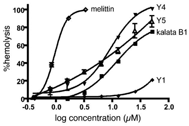
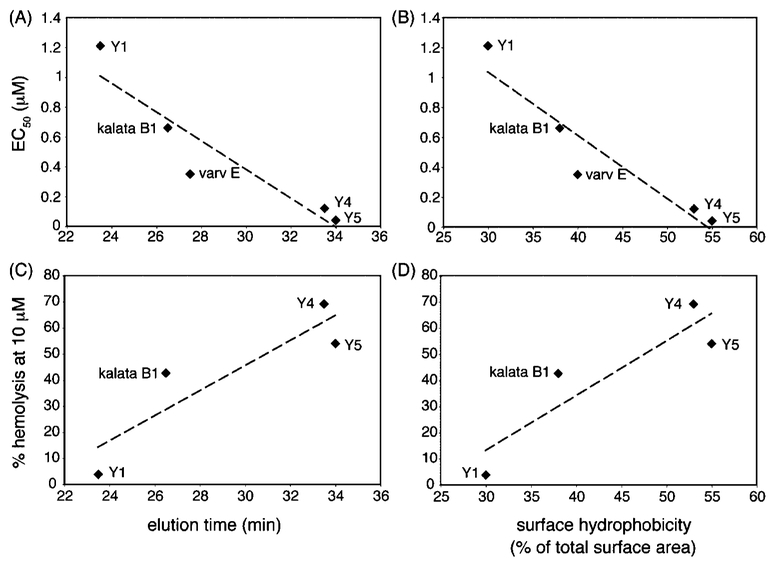
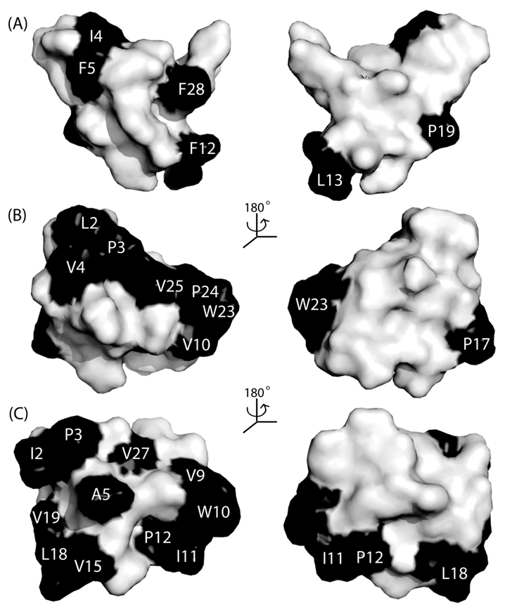
Cyclotides are macrocyclic plant peptides characterized by a knotted arrangement of three disulfide bonds. They display a range of interesting bioactivities, including anti-HIV and insecticidal activities. More than 100 different cyclotides have been isolated from two phylogenetically distant plant families, the Rubiaceae and Violaceae. In this study we have characterized the cyclotides from Viola yedoensis, an important Chinese herb from the Violaceae family that has been reported to contain potential anti-HIV agents. From V. yedoensis five new and three known cyclotides were identified and shown to have anti-HIV activity. The most active of these is cycloviolacin Y5, which is one of the most potent of all cyclotides tested so far using in vitro XTT-based anti-HIV assays. Cycloviolacin Y5 is the most hydrophobic of the cyclotides from V. yedoensis. We show that there is a positive correlation between the hydrophobicity and the anti-HIV activity of the new cyclotides and that this trend tracks with their ability to disrupt membranes, as judged from hemolytic assays on human erythrocytes.
Figures
Similar articles
-
Cycloviolacin H4, a hydrophobic cyclotide from Viola hederaceae.J Nat Prod. 2006 Jan;69(1):23-8. doi: 10.1021/np050317i. J Nat Prod. 2006. PMID: 16441062
-
Isolation and characterization of cytotoxic cyclotides from Viola tricolor.Peptides. 2010 Aug;31(8):1434-40. doi: 10.1016/j.peptides.2010.05.004. Epub 2010 May 16. Peptides. 2010. PMID: 20580652
-
Isolation and characterization of cytotoxic cyclotides from Viola philippica.Peptides. 2011 Aug;32(8):1719-23. doi: 10.1016/j.peptides.2011.06.016. Epub 2011 Jun 23. Peptides. 2011. PMID: 21723349
-
An overview of acyclotides: Past, present and future.Phytochemistry. 2020 Feb;170:112215. doi: 10.1016/j.phytochem.2019.112215. Epub 2019 Dec 4. Phytochemistry. 2020. PMID: 31812106 Review.
-
Potential therapeutic applications of the cyclotides and related cystine knot mini-proteins.Expert Opin Investig Drugs. 2007 May;16(5):595-604. doi: 10.1517/13543784.16.5.595. Expert Opin Investig Drugs. 2007. PMID: 17461734 Review.
Cited by
-
Fighting nature with nature: antiviral compounds that target retroviruses.Arch Microbiol. 2024 Feb 28;206(3):130. doi: 10.1007/s00203-024-03846-3. Arch Microbiol. 2024. PMID: 38416180 Review.
-
A Synthetic mirror image of kalata B1 reveals that cyclotide activity is independent of a protein receptor.Chembiochem. 2011 Nov 4;12(16):2456-62. doi: 10.1002/cbic.201100450. Epub 2011 Sep 16. Chembiochem. 2011. PMID: 21928440 Free PMC article.
-
Anti-inflammatory effects of Viola yedoensis and the application of cell extraction methods for investigating bioactive constituents in macrophages.BMC Complement Altern Med. 2016 Jun 14;16:180. doi: 10.1186/s12906-016-1142-9. BMC Complement Altern Med. 2016. PMID: 27301877 Free PMC article.
-
Dynamic scenario of membrane binding process of kalata b1.PLoS One. 2014 Dec 4;9(12):e114473. doi: 10.1371/journal.pone.0114473. eCollection 2014. PLoS One. 2014. PMID: 25473840 Free PMC article.
-
A Systematic Review of Phytochemistry, Nutritional Composition, and Pharmacologic Application of Species of the Genus Viola in Noncommunicable Diseases (NCDs).Evid Based Complement Alternat Med. 2023 Oct 31;2023:5406039. doi: 10.1155/2023/5406039. eCollection 2023. Evid Based Complement Alternat Med. 2023. PMID: 37941895 Free PMC article. Review.
References
-
- Craik DJ; Daly NL; Bond T; Waine CJ Mol. Biol 1999, 294, 1327–1336. - PubMed
-
- Craik DJ; Daly NL; Mulvenna J; Plan MR; Trabi M Curr. Protein Pept. Sci 2004, 5, 297–315. - PubMed
-
- Goransson U; Luijendijk T; Johansson S; Bohlin L; Claeson PJ Nat. Prod 1999, 62, 283–286. - PubMed
-
- Gustafson KR; Walton LK; Sowder RCJ; Johnson DG; Pannell LK; Cardellina JH II; Boyd MR J. Nat. Prod 2000, 63, 176–178. - PubMed
MeSH terms
Substances
Grants and funding
LinkOut - more resources
Full Text Sources
Other Literature Sources
Medical
