. 2008 Jan 8;47(1):479-89.
doi: 10.1021/bi7013882. Epub 2007 Dec 15.

S2 deletion variants of human PRL receptors demonstrate that extracellular domain conformation can alter conformation of the intracellular signaling domain

Affiliations

S2 deletion variants of human PRL receptors demonstrate that extracellular domain conformation can alter conformation of the intracellular signaling domain

Dunyong Tan et al. Biochemistry. .

Abstract

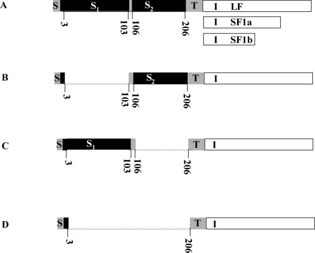
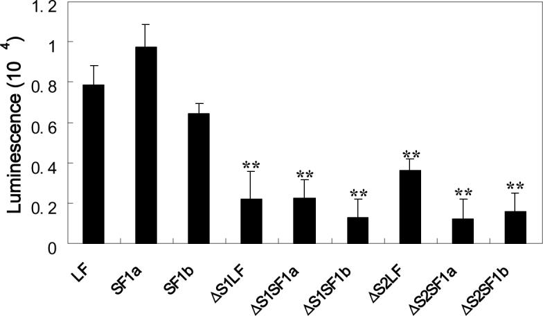
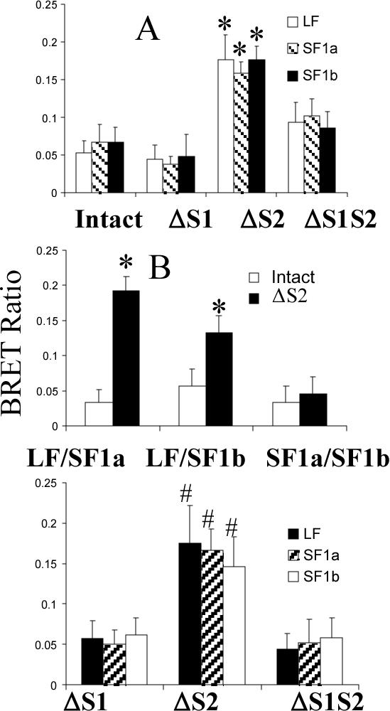
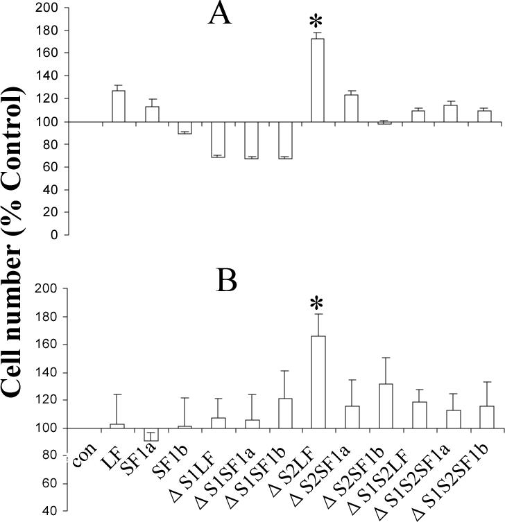
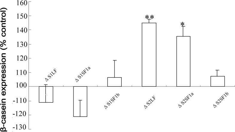
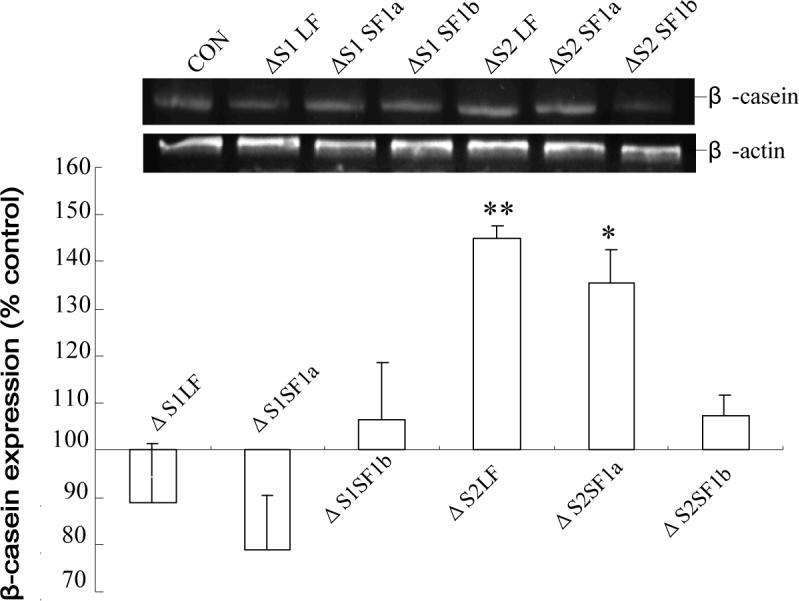
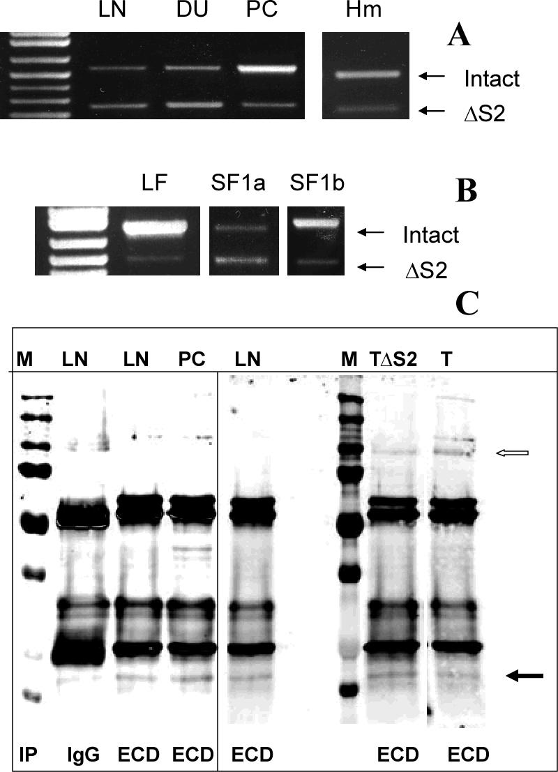
Using spacers between the C-termini of the long (LF) or short (SF) human prolactin receptors and luciferase/GFP such that bioluminescence resonance energy transfer (BRET) occurred minimally in intact versions of these receptors in the absence of ligand, we have monitored the BRET signal after deletion of regions of the extracellular domain (ECD). Deletion of S2 produced ligand-independent BRET for only those pairings normally occurring in the presence of ligand with the intact receptor. Deletion of the similarly sized S1, or S1 plus S2, produced no ligand-independent or -dependent BRET. When deleted receptors were transfected into human breast (T47D) or prostate (DU145) cancer cells incubated in the absence of added prolactin (PRL) and presence of anti-PRL, expression of the DeltaS2LF resulted in increased cell number, whereas expression of the intact receptor did not. When endogenous beta-casein expression was examined in T47D cells, the DeltaS2LF and DeltaS2F1a both showed ligand-independent activation of transcription, again not duplicated by the intact receptor. Paired with evidence in the literature for predimerization of PRLRs, these results demonstrate that altered ECD conformation, and not just a change in bulk, produces altered conformation of the intracellular signaling region of the receptors, supporting the concept that ligand binding to the ECD of intact predimerized receptors could initiate signaling. In addition, the current work supports a dual proliferative and differentiative role for the LF receptor, but only a differentiative role for the SF1a receptor. Naturally occurring DeltaS2 PRL receptors (PRLR) were also found in normal and cancerous human cells. This additionally suggests a heretofore unappreciated ligand-independent role for PRLRs.


Figures

Fig 1
Fig 1. Diagram illustrating the different versions of the PRLR used
A, intact human LF PRLR with the shorter intracellular regions of SF1a and SF1b illustrated. The transmembrane and ECD regions are the same for all three; B, S1- deleted; C, S2-deleted and D, S1- and S2-deleted The dotted line illustrates the part deleted and the numbers given are the amino acids in the mature sequence. S, signal peptide; S1, subdomain 1; S2, subdomain 2, of the extracellular domain; T, transmembrane; I, intracellular part of long form (LF), short form (SF) 1a and SF1b.
Fig 2
Fig 2. Confocal images demonstrating expression, localization (A) and spectral properties (B) of the GFP2 and/or Rluc-tagged deleted human PRLRs
HEK 293 cells transiently transfected with plasmids containing cDNA coding for the receptors tagged with GFP2 were examined by confocal microscopy after 48h. bar,10μm; white arrowhead, localization of the tagged receptors to the region of the plasma membrane. For spectral scanning, GFP2 was excited at 405nm. Bioluminescence generated by Rluc was detected in the presence of substrate, DeepBlueC (5 μM), with blockade of the external excitation light source. Only the spectra for the ΔS1 versions are shown by way of example, but all spectra were unaltered by attachment to the various receptors. GFP2N1 is the GFP2 expressing vector without any receptor, and RLuc1N1 is the luciferase expressing vector without receptor. The relative expression of each receptor can be appreciated by comparing the amount of fluorescence or luciferase activity. Relative expression levels were the same for the intact and deleted versions of the receptor.
Fig 3
Fig 3. Biological activity of PRL-Gluc and the relative ability of the intact and deleted PRLRs on HEK293 cells to bind and internalize PRL-Gluc
Panel A compares diluted conditioned medium with unmodified human PRL. PRL-Gluc was biologically active, reaching the same maximum as unmodified PRL. OD 492 nm is absorbance in the MTS assay and is representative of relative cell number. A dilution of conditioned medium with bioactivity equivalent to 50ng/ml unmodified PRL was used for the studies in panel B. After a 3h incubation at 37°C, the cells were washed three times and cell-associated luminescence was determined. Non-specific binding/uptake (binding/uptake in the presence of excess unmodified PRL for each receptor type) was subtracted from each result. ** significantly different from their intact counterpart with p<0.01.
Fig 4
Fig 4. Bioluminescence resonance energy transfer with intact and variously deleted receptors and combinations of receptors in the absence (A and B) and presence (C) of PRL
HEK 293 cells were transfected with equal quantities of GFP2- or luc-tagged receptors and 48h later were subjected to BRET analysis initiated by the addition of substrate. For panel B, the same result was obtained regardless of which receptor in the pair was tagged with GFP or luc. Results in all three panels are the mean ± SEM derived from experiments in which all variables from all panels were concurrently analyzed. Energy transfer is given as the BRET ratio (Emission500nm-520nm-background500nm-520nm)/(Emission385nm-420nm-background 385nm-420nm). BRET signals obtained from nontransfected cells were considered background in this equation. *, significantly different from the intact version with p<0.05; #, significantly different from ΔS1 and ΔS1S2 (p<0.05) and not different from ΔS2 in panel A.
Fig 5
Fig 5. Effect of intact and deleted PRLRs on the growth of human breast cancer T47D cells (A) and human prostate cancer DU145 cells (B) in the absence of added PRL
Receptor constructs were transfected into either T47D or DU145 cells. Forty eight hours post transfection, the medium with serum was refreshed. After a further 24h, the medium was changed to serum-free DMEM to conduct the MTS assay. Data are expressed as a percent of the control to illustrate the change induced. The data are not corrected for either transfection efficiency or expression efficiency for reasons discussed in the text. The same overall result was obtained when anti-PRL was added to the incubation, although cell number was decreased in each incubation indicative of an effect of the antibody on autocrine PRL. *, significantly different from the intact counterpart with p<0.05.
Fig 6
Fig 6. Effect of the ΔS1 and ΔS2 receptors on endogenous β-casein gene expression in T47D cells
Forty eight hours post transfection, the cells were subjected to a further 24 h incubation in the absence of added PRL and then RT-PCR for β-casein was performed. Data are expressed relative to expression in non-transfected cells (which was the same as that in cells expressing the doubly deleted S1S2 receptor) to illustrate the changes observed. The data are not corrected for either transfection or expression efficiency. The same overall result was obtained in the presence of anti-PRL. *, significantly different from both the non-transfected and ΔS1S2-transfected cells with p<0.05.
Fig 7
Fig 7. Natural expression of ΔS2 PRLRs
(A) Amplicons resulting from the expression of both intact and ΔS2 PRLR were observed in human LNCaP (LN), DU145 (DU), PC3 (PC) and microvessel human endothelial (Hm) cells and their identities were confirmed by sequencing. (B) Use of form-specific reverse primers demonstrated the presence of a ΔS2 variant of each receptor in all cells, and PC3 cells are illustrated by way of example. Again, the identities of the amplicons were verified by sequencing. Note that different numbers of cycles were required to demonstrate this for each receptor isoform and so the panels cannot be compared in terms of relative abundance (LF and SF1a, 35 cycles; SF1b, 40 cycles). (C) After immunoprecipitation, ΔS2 versions of SF1b are illustrated (closed arrow) as detected by Western blot (using the same anti-ECD antibody) in PC3, LNCaP and T47D (T) cells. Also illustrated is co-migration of immunoprecipitated ΔS2SF1b stably expressed under the control of tetracycline in T47D cells (TΔS2). M, molecular mass markers; IP, immunoprecipitated with control isotype-matched antibody (IgG) or antibody against the extracellular domain (ECD); open arrow marks a LF of the receptor. The vertical line separates samples run on different gels. The sample from normal T47D cells (T) was run on the same gel, but the lane was cut and pasted to be next to the TΔS2 cells to eliminate distracting additional experiments.

Similar articles

Cited by

References

    1. Nevalainen MT, Valve EM, Ingleton PM, Nurmi M, Martikainen PM, Harkonen PL. Prolactin and prolactin receptors are expressed and functioning in human prostate. J. Clin. Invest. 1997;99:618–627. - PMC - PubMed
    1. Neville MC, McFadden TB, Forsyth I. Hormonal regulation of mammary differentiation and milk secretion. J. Mammary Gland Biol. Neoplasia. 2002;7:49–66. - PubMed
    1. Goffin V, Binart N, Touraine P, Kelly PA. Prolactin: the new biology of an old hormone. Ann. Rev. Physiol. 2002;64:47–67. - PubMed
    1. Hu Z, Zhuang L, Maria L, Dufau ML. Multiple and Tissue-specific Promoter Control of Gonadal and Non-gonadal Prolactin Receptor Gene Expression. J. Biol. Chem. 1996;271:10242–10246. - PubMed
    1. Nagano M, Kelly PA. Tissue distribution and regulation of rat prolactin receptor gene expression. Quantitative analysis by polymerase chain reaction. J. Biol. Chem. 1994;269:13337–13345. - PubMed

Publication types

MeSH terms