

Structure and function of PA4872 from Pseudomonas aeruginosa, a novel class of oxaloacetate decarboxylase from the PEP mutase/isocitrate lyase superfamily
- PMID: 18081320
- PMCID: PMC2892964
- DOI: 10.1021/bi701954p
Structure and function of PA4872 from Pseudomonas aeruginosa, a novel class of oxaloacetate decarboxylase from the PEP mutase/isocitrate lyase superfamily
Abstract
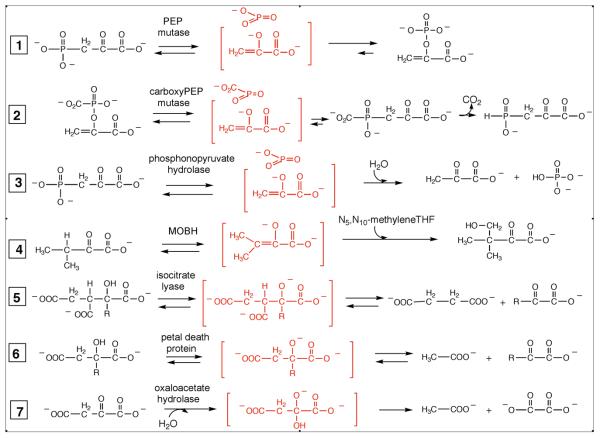
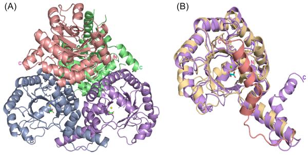
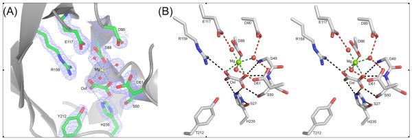
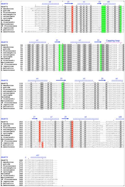
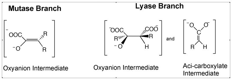
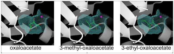
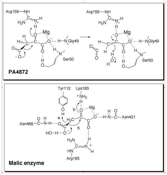
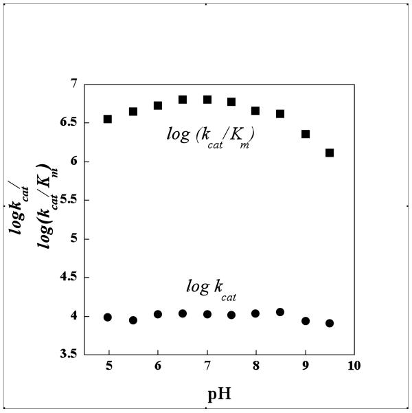
Pseudomonas aeruginosa PA4872 was identified by sequence analysis as a structurally and functionally novel member of the PEP mutase/isocitrate lyase superfamily and therefore targeted for investigation. Substrate screens ruled out overlap with known catalytic functions of superfamily members. The crystal structure of PA4872 in complex with oxalate (a stable analogue of the shared family alpha-oxyanion carboxylate intermediate/transition state) and Mg2+ was determined at 1.9 A resolution. As with other PEP mutase/isocitrate lyase superfamily members, the protein assembles into a dimer of dimers with each subunit adopting an alpha/beta barrel fold and two subunits swapping their barrel's C-terminal alpha-helices. Mg2+ and oxalate bind in the same manner as observed with other superfamily members. The active site gating loop, known to play a catalytic role in the PEP mutase and lyase branches of the superfamily, adopts an open conformation. The Nepsilon of His235, an invariant residue in the PA4872 sequence family, is oriented toward a C(2) oxygen of oxalate analogous to the C(3) of a pyruvyl moiety. Deuterium exchange into alpha-oxocarboxylate-containing compounds was confirmed by 1H NMR spectroscopy. Having ruled out known activities, the involvement of a pyruvate enolate intermediate suggested a decarboxylase activity of an alpha-oxocarboxylate substrate. Enzymatic assays led to the discovery that PA4872 decarboxylates oxaloacetate (kcat = 7500 s(-1) and Km = 2.2 mM) and 3-methyloxaloacetate (kcat = 250 s(-1) and Km = 0.63 mM). Genome context of the fourteen sequence family members indicates that the enzyme is used by select group of Gram-negative bacteria to maintain cellular concentrations of bicarbonate and pyruvate; however the decarboxylation activity cannot be attributed to a pathway common to the various bacterial species.
Figures

Similar articles
-
Structure and kinetics of phosphonopyruvate hydrolase from Variovorax sp. Pal2: new insight into the divergence of catalysis within the PEP mutase/isocitrate lyase superfamily.Biochemistry. 2006 Sep 26;45(38):11491-504. doi: 10.1021/bi061208l. Biochemistry. 2006. PMID: 16981709
-
Helix swapping between two alpha/beta barrels: crystal structure of phosphoenolpyruvate mutase with bound Mg(2+)-oxalate.Structure. 1999 May;7(5):539-48. doi: 10.1016/s0969-2126(99)80070-7. Structure. 1999. PMID: 10378273
-
The crystal structure and active site location of isocitrate lyase from the fungus Aspergillus nidulans.Structure. 2000 Apr 15;8(4):349-62. doi: 10.1016/s0969-2126(00)00117-9. Structure. 2000. PMID: 10801489
-
Coupling mechanism of the oxaloacetate decarboxylase Na(+) pump.Biochim Biophys Acta. 2001 May 1;1505(1):1-14. doi: 10.1016/s0005-2728(00)00272-3. Biochim Biophys Acta. 2001. PMID: 11248184 Review.
-
Pericyclic reactions catalyzed by chorismate-utilizing enzymes.Biochemistry. 2011 Sep 6;50(35):7476-83. doi: 10.1021/bi2009739. Epub 2011 Aug 12. Biochemistry. 2011. PMID: 21823653 Free PMC article. Review.
Cited by
-
Meta-analysis highlights the key drought responsive genes in genes: PEPC and TaSAG7 are hubs response networks.J Genet Eng Biotechnol. 2022 Sep 2;20(1):127. doi: 10.1186/s43141-022-00395-4. J Genet Eng Biotechnol. 2022. PMID: 36053361 Free PMC article.
-
The PEP-pyruvate-oxaloacetate node: variation at the heart of metabolism.FEMS Microbiol Rev. 2021 May 5;45(3):fuaa061. doi: 10.1093/femsre/fuaa061. FEMS Microbiol Rev. 2021. PMID: 33289792 Free PMC article.
-
Structure of oxalacetate acetylhydrolase, a virulence factor of the chestnut blight fungus.J Biol Chem. 2010 Aug 20;285(34):26685-96. doi: 10.1074/jbc.M110.117804. Epub 2010 Jun 17. J Biol Chem. 2010. PMID: 20558740 Free PMC article.
-
Structural and kinetic characterization of 4-hydroxy-4-methyl-2-oxoglutarate/4-carboxy-4-hydroxy-2-oxoadipate aldolase, a protocatechuate degradation enzyme evolutionarily convergent with the HpaI and DmpG pyruvate aldolases.J Biol Chem. 2010 Nov 19;285(47):36608-15. doi: 10.1074/jbc.M110.159509. Epub 2010 Sep 15. J Biol Chem. 2010. PMID: 20843800 Free PMC article.
-
The role of biotin and oxamate in the carboxyltransferase reaction of pyruvate carboxylase.Arch Biochem Biophys. 2014 Nov 15;562:70-9. doi: 10.1016/j.abb.2014.08.008. Epub 2014 Aug 23. Arch Biochem Biophys. 2014. PMID: 25157442 Free PMC article.
References
-
- Liu S, Lu Z, Han Y, Melamud E, Dunaway-Mariano D, Herzberg O. Crystal structures of 2-methylisocitrate lyase in complex with product and with isocitrate inhibitor provide insight into lyase substrate specificity, catalysis and evolution. Biochemistry. 2005;44:2949–2962. - PubMed
-
- Britton K, Langridge S, Baker PJ, Weeradechapon K, Sedelnikova SE, De Lucas JR, Rice DW, Turner G. The crystal structure and active site location of isocitrate lyase from the fungus Aspergillus nidulans. Structure. 2000;8:349–362. - PubMed
-
- Britton KL, Abeysinghe IS, Baker PJ, Barynin V, Diehl P, Langridge SJ, McFadden BA, Sedelnikova SE, Stillman TJ, Weeradechapon K, Rice DW. The structure and domain organization of Escherichia coli isocitrate lyase. Acta Crystallogr D Biol Crystallogr. 2001;57:1209–1218. - PubMed
-
- Chaudhuri BN, Sawaya MR, Kim CY, Waldo GS, Park MS, Terwilliger TC, Yeates TO. The crystal structure of the first enzyme in the pantothenate biosynthetic pathway, ketopantoate hydroxymethyltransferase, from M tuberculosis. Structure. 2003;11:753–764. - PubMed
-
- Chen CC, Han Y, Niu W, Kulakova AN, Howard A, Quinn JP, Dunaway-Mariano D, Herzberg O. Structure and kinetics of phosphonopyruvate hydrolase from Variovorax sp. Pal2: new insight into the divergence of catalysis within the PEP mutase/isocitrate lyase superfamily. Biochemistry. 2006;45:11491–11504. - PubMed
Publication types
MeSH terms
Substances
Associated data
- Actions
Grants and funding
LinkOut - more resources
Full Text Sources
Other Literature Sources
Chemical Information
Molecular Biology Databases
Research Materials
Miscellaneous
