

Review
doi: 10.1111/j.1365-2249.2007.03552.x.
Translational mini-review series on complement factor H: genetics and disease associations of human complement factor H
Affiliations
- PMID: 18081690
- PMCID: PMC2276932
- DOI: 10.1111/j.1365-2249.2007.03552.x
Item in Clipboard
Review
Translational mini-review series on complement factor H: genetics and disease associations of human complement factor H
Clin Exp Immunol.
2008 Jan.
Abstract
Factor H is an abundant plasma glycoprotein that plays a critical role in the regulation of the complement system in plasma and in the protection of host cells and tissues from damage by complement activation. Several recent studies have described the association of genetic variations of the complement factor H gene (CFH) with atypical haemolytic uraemic syndrome (aHUS), age-related macular degeneration (AMD) and membranoproliferative glomerulonephritis (MPGN). This review summarizes our current knowledge of CFH genetics and examines the CFH genotype-phenotype correlations that are helping to understand the molecular basis underlying these renal and ocular pathologies.
Figures
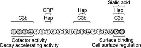
Functional domains in complement factor H. Factor H has three C3b-binding sites, short consensus repeats (SCR)1–4, SCR12–14 and SCR19–20, respectively [–13]. Similarly, a total of three separate binding sites for heparin and sialic acid have been identified in SCR7, SCR13 and SCR19–20, respectively [–17]. The critical sites for co-factor activity/decay accelerating activity and cell surface regulation at the N- and C-termini, respectively, are indicated. In addition to C3b and polyanion binding sites, there are other domains in factor H that have been shown to interact with plasma proteins or with micoorganisms and that are interesting because of their potential relevance in pathology. In this regard, it has been shown that factor H binds to C-reactive protein (CRP) which may help to counteract and inhibit the CRP-dependent alternative pathway activation induced by damaged tissue [18,19]. The heparin- and CRP-binding sites in SCR7 are overlapping sites in which one substrate inhibits the binding of the others [20].
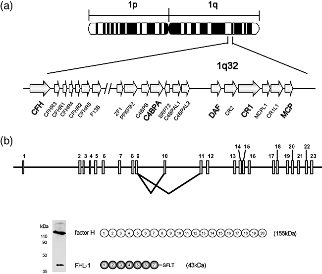
Chromosomal location and structure of the factor H gene. (a) The human regulator of complement activation (RCA) gene cluster in 1q32. The human RCA gene cluster spans a total of 21·45 cM and includes more than 60 genes of which 15 are complement-related genes. All of the complement-related genes are arranged in tandem within two groups. The two groupings are a telomeric 900 kb-long DNA segment which contains the C4BPB, C4BPA, C4BPAL1, C4BPAL2, DAF, CR2, CR1, MCPL1, CR1L1 and MCP genes and a centromeric 650 kb-long DNA segment that contains CFH, CFHR3, CFHR1, CFHR4, CFHR2 and CFHR5, as well as the gene coding for the B subunit of the coagulation factor XIII, F13B. These two gene groups are separated by 14·59 cM, a large amount of DNA-containing genes that are unrelated to complement and that have very diverse functions [38]. It is generally accepted that these complement regulatory genes share a common ancestor from which they originated by multiple events of gene duplication. (b) Structure of CFH, showing a diagram of the 23 exons and the two alternative splicing products of the CFH gene. Exon 10 does not contribute to the factor H transcript but it is utilized for the FHL-1 molecule. The figure also shows a Western blot, using a monoclonal antibody (35H9) that recognizes both factor H and FHL-1, to illustrate the relative amounts of these proteins in normal human plasma.
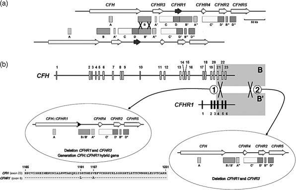
The complement factor H gene (CFH)–CFHR1–5 gene subregion of the regulator of complement activation (RCA) gene cluster. (a) Genomic organization of the CFH and CFHR1–5 genes and location of low copy number repeats. Arrows represent the genes with their names above. The boxes underneath indicate the sequence repeats. Low copy repeats are named with the same letter (i.e. A, A′, A′′). A grey colour-code is used to identify the different repeats. The figure shows two chromosomes aligned by the B and B′ repeats to illustrate the CFH–CFHR1 genomic that occurred through non-homologous recombination between a 23 kb-long repeat region in the 3′ end of the CFH and CFHR1 genes, labelled B and B′, respectively. (b) Deletion of the CFHR1 and CFHR3 genes and generation of a CFH::CFHR1 hybrid gene. The rearrangement marked 2 is relatively common in multiple African and European populations. This rearrangement involves non-homologous recombination between the B and B′ homologous regions downstream of the CFH and CFHR1 genes and results in no sequence-modification of the CFH gene. A second rearrangement (labelled 1) is much less frequent and found associated exclusively with atypical haemolytic uraemic syndrome (aHUS). Non-homologous cross-over in this case occurred between the B and B′ homologous regions in intron 21 of CFH and intron 4 of CFHR1 and results in generation of a hybrid CFH::CFHR1 gene. The hybrid gene consists of the first 21 exons of CFH [encoding SCRs 1–18 of CFH] and the last two exons of CFHR1 (encoding SCR4 and 5 of CFHR1) [44]. Amino acid sequences of CFH exon 23 and CFHR1 exon 6 are aligned to illustrate two amino acid differences between them (S1191L/V1197A) that are present in the protein product of the hybrid gene. These amino acid changes in SCR20 are associated with aHUS and are identical to those present in the CFH mutant protein, also associated with aHUS, that generates by gene conversion [45].
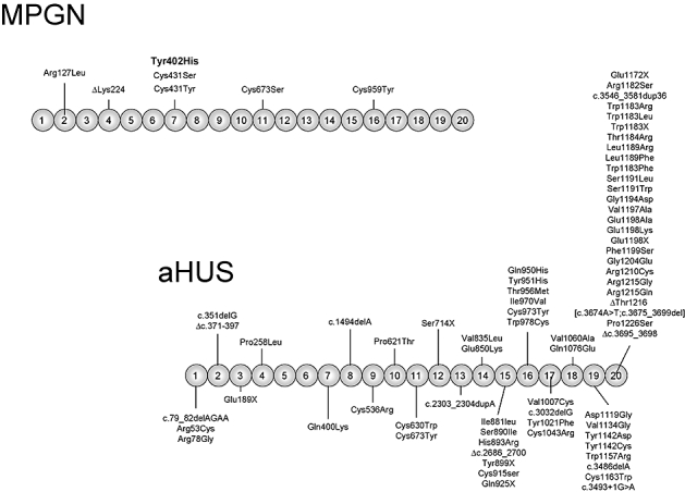
Complement factor H gene (CFH) mutations in MPGN and atypical haemolytic uraemic syndrome (aHUS) patients. The figure shows a diagram of the structure of human factor H with the 20 SCRs. The location of the missense mutations characterized thus far in membranoproliferative glomerulonephritis (MPGN) and aHUS patients is indicated. The position of the Tyr402His polymorphisms associated strongly with predisposition to AMD is highlighted in bold. Note that mutations associated with aHUS are clustered in the C-terminus, the region of factor H that is critical for the control of C3b deposited on cell surfaces.
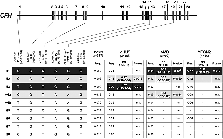
Complement factor H gene (CFH) haplotypes and their association with disease. Schematic illustration of the CFH exon structure showing the location of the six single nucleotide polymorphisms (SNPs) included in these studies. These SNPs represent a minimal informative set for genetic variation within the CFH gene. Haplotype frequencies in the control and patient cohorts were estimated using the expectation maximization (EM) algorithm implemented by the SNPStats software (available on-line at: http://bioinfo.iconcologia.net/SNPstats ). CFH haplotypes with a frequency > 1% are shown. The frequency of each CFH haplotype was compared between the controls and the atypical haemolytic uraemic syndrome, age-related macular degeneration and membranoproliferative glomerulonephritis type II cohorts and the P-values and the odds ratios (OR) were calculated. Risk haplotypes are shaded black, while protective haplotypes are shaded in grey. P-values were derived using the two-sided Fisher's exact test. OR and 95% confidence intervals are shown. The nucleotide and amino acid numbering are referred to the translation start site (A in ATG is +1; Met is +1) as recommended by the Human Genome Variation Society. This figure is an updated version of that published in Pickering et al. [66].
References
-
- Law SKA, Reid KBM. Complement. 2. Oxford: IRL Press; 1995.
Publication types
MeSH terms
Substances
LinkOut - more resources
Full Text Sources
Other Literature Sources
Medical
Miscellaneous
