

Review
doi: 10.1016/j.conb.2007.11.004.
Epub 2007 Dec 21.
Circuit reconstruction tools today
Affiliations
- PMID: 18082394
- PMCID: PMC2693015
- DOI: 10.1016/j.conb.2007.11.004
Item in Clipboard
Review
Circuit reconstruction tools today
Curr Opin Neurobiol.
2007 Oct.
Abstract
To understand how a brain processes information, we must understand the structure of its neural circuits-especially circuit interconnection topologies and the cell and synapse molecular architectures that determine circuit-signaling dynamics. Our information on these key aspects of neural circuit structure has remained incomplete and fragmentary, however, because of limitations of the best available imaging methods. Now, new transgenic tool mice and new image acquisition tools appear poised to permit very significant advances in our abilities to reconstruct circuit connection topologies and molecular architectures.
Figures
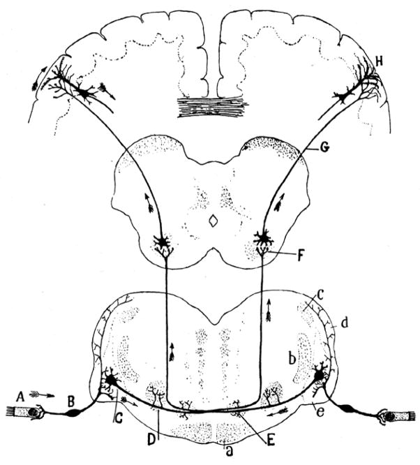
Drawings like this built the foundations of modern neuroscience, establishing the idea that brains process information and generate behavior as a result of the conduction of signals from cell to cell through anatomically defined circuits. Arrows in Ramon y Cajal’s india-ink reconstruction of the auditory pathway (Ref [1], fig. I-26) indicate information flow, from auditory hair cells (A) through the ventral cochlear nucleus (C) and the inferior colliculus (F) to cortical pyramidal cells (H), and then corticofugally to control behavior, via the axonal projections of cortical pyramidal cells. Like all subsequent reconstructions of brain circuitry, this early reconstruction is far from complete.
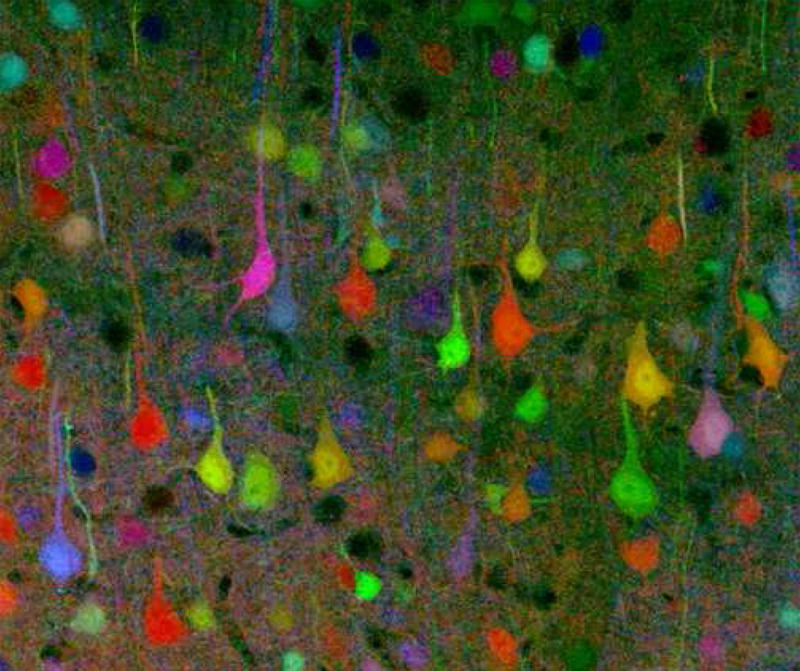
Reference introduces a powerful new molecular genetic method for generating mice in which a large fraction of neurons express fluorescence tags drawn randomly from a large combinatorial color palette. (This image provided courtesy of Prof. Jeff Lichtman on behalf of all authors of Ref. 12). Such “color coded” brain cells promise new solutions to formerly intractable problems with resolving closely packed neurons and tracing their axons and dendrites reliably over long distances. It seems unlikely that neural information processing will ever be understood without solving such problems and reconstructing circuits in far more detail that presently possible.
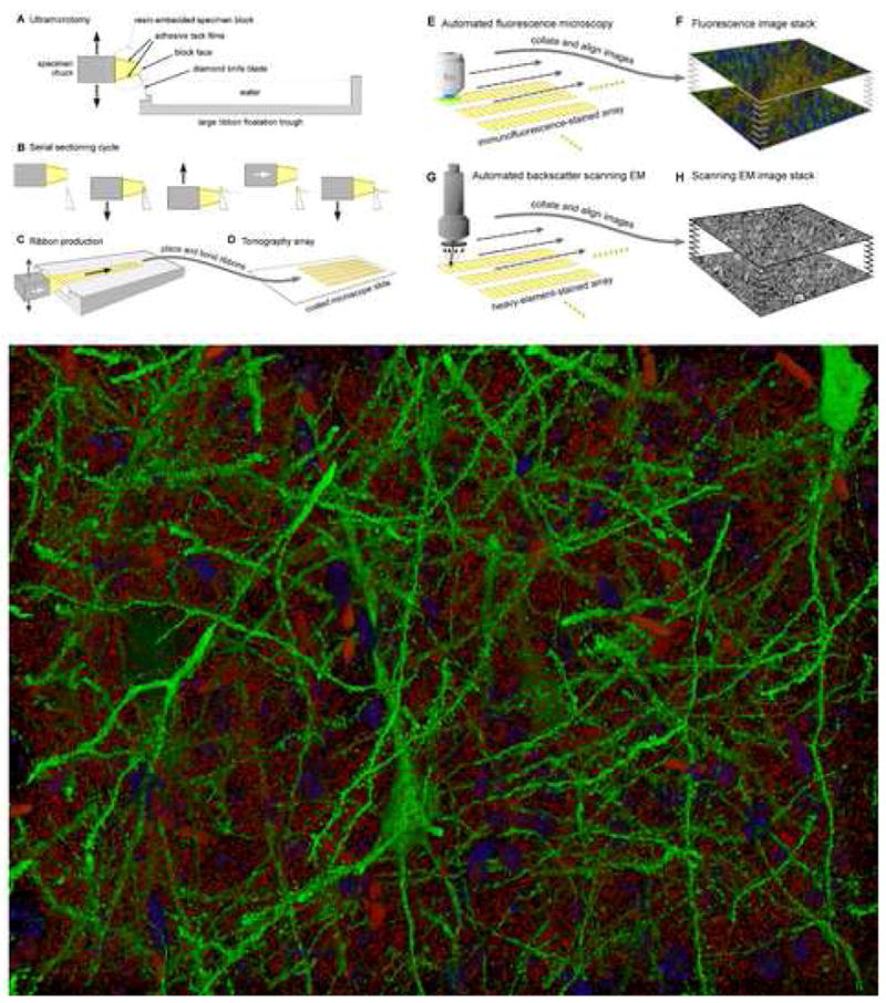
The reconstruction of neural circuits will benefit from complementing the molecular discrimination of immunofluorescence imaging with the structural precision of electron microscopy. A. - D. Tomography array production. An ultramicrotome (A) cuts a resin-embedded specimen into sections 50–200nm thick by motion (B) against a diamond knife blade. Adhesive block coatings cause serial sections to form continuous ribbons (C), which are transferred and bonded to a glass array slide (D). E., F. Immunofluorescence imaging. Array slides are immunostained and imaged using an automated fluorescence microscope (E). Resulting two-dimensional images are then aligned to form a three-dimensional image stack (F). Repeated cycles of immuno-staining, imaging and antibody elution allow multiplexing of very large numbers of immunofluorescence channels [15]. G., H. Electron-microscopy. After immunofluorescence imaging, arrays can be re-stained for imaging by SEM (G), providing unique opportunities to tap complementary strengths of immuno-fluorescence and electron microscopic volume imaging (H). I. Volume rendering of an array tomographic immunofluorescence image of a subset of layer 5 pyramidal cells (green) and putative synapses (anti-synapsin-I puncta, red) in a 180×140×30 um volume of mouse whisker barrel. Blue objects are DAPI stained nuclei of otherwise unstained cells. The larger red objects are erythrocytes within capillaries. The specimen is from a Line H Thy-1-YFP mouse [6]. See reference for examples of conjugate immunofluorescence and SEM array tomography.
References
-
- Ramon y Cajal S. In: Histology of the Nervous System of Man and Vertebrates. Swanson N, Swanson L, editors. Oxford Press; 1995. Summarizes the work that first placed the concept of information processing by neural circuit structures on a firm anatomical foundation.**
-
- Nelson SB, Hempel C, Sugino K. Probing the transcriptome of neuronal cell types. Curr Opin Neurobiol. 2006 Oct;16(5):571–6. Epub 2006 Sep 7. - PubMed
-
- Molyneaux BJ, Arlotta P, Menezes JR, Macklis JD. Neuronal subtype specification in the cerebral cortex. Nat Rev Neurosci. 2007;6:427–437. - PubMed
-
- Lein ES, et al. Genome-wide atlas of gene expression in the adult mouse brain. Nature. 2007;445:168–176. - PubMed
Publication types
MeSH terms
Grants and funding
LinkOut - more resources
Full Text Sources
