. 2007 Dec 14;28(5):823-37.
doi: 10.1016/j.molcel.2007.11.012.

PHD domain-mediated E3 ligase activity directs intramolecular sumoylation of an adjacent bromodomain required for gene silencing

Affiliations

PHD domain-mediated E3 ligase activity directs intramolecular sumoylation of an adjacent bromodomain required for gene silencing

Alexey V Ivanov et al. Mol Cell. .

Abstract

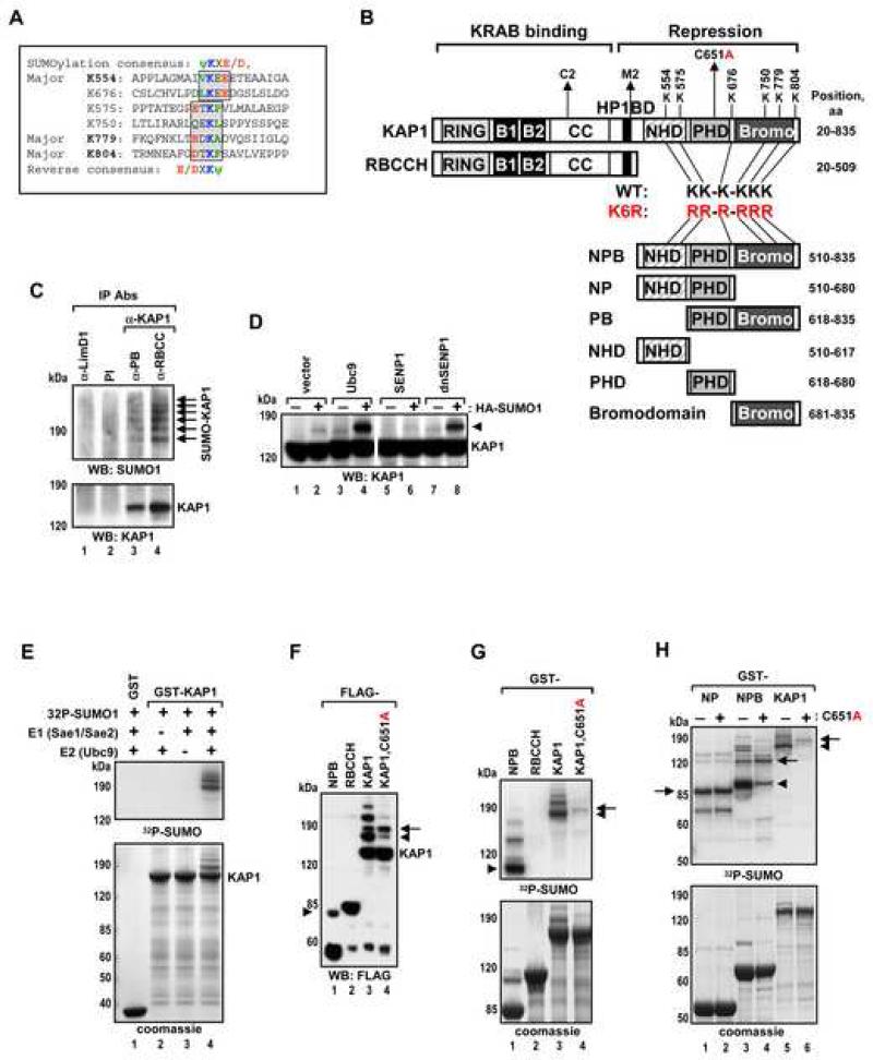
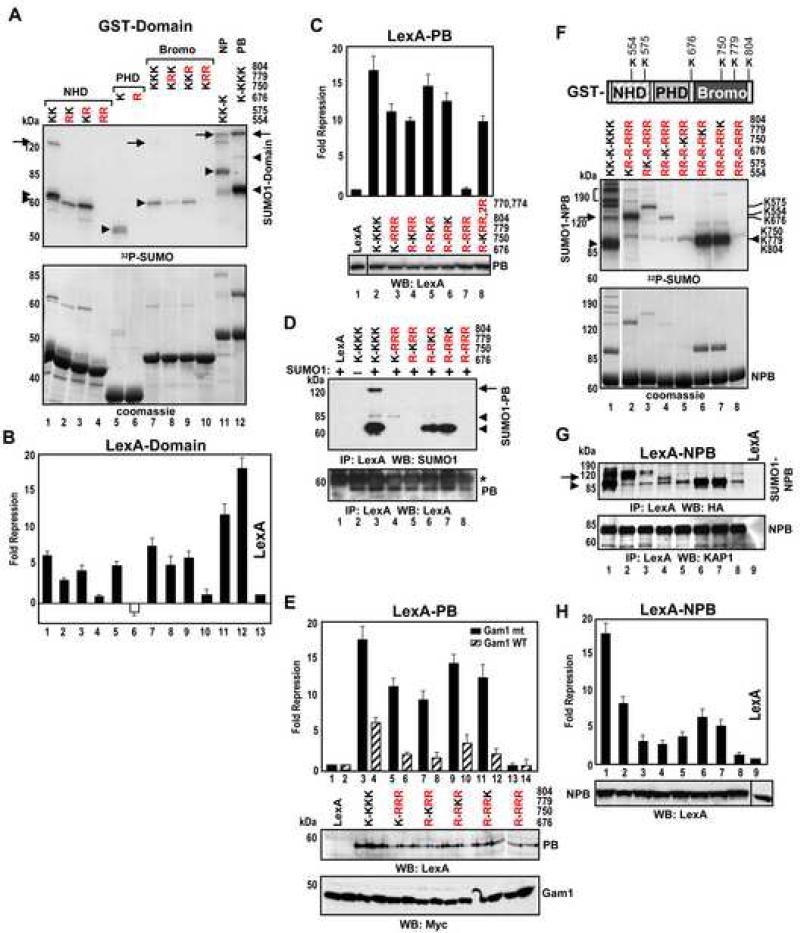
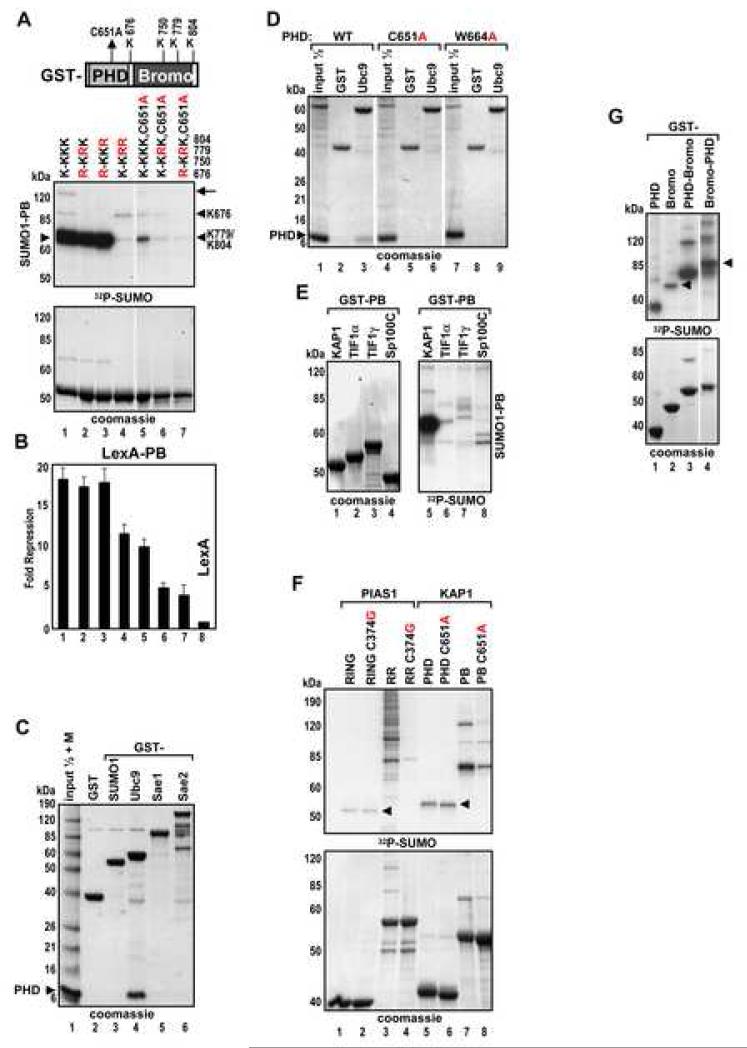
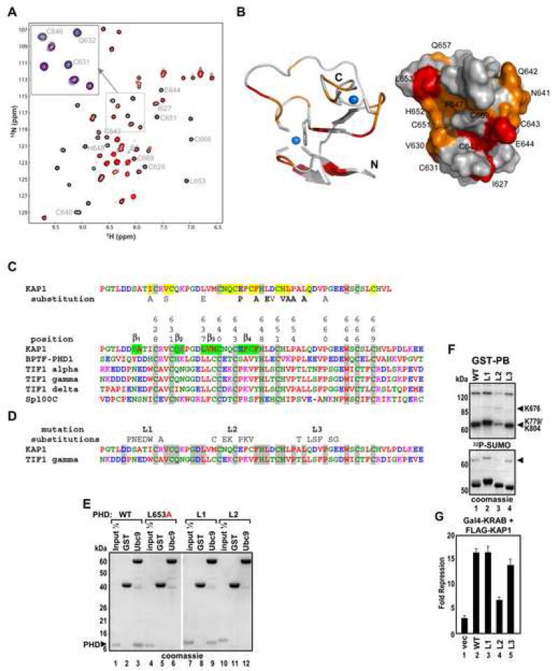
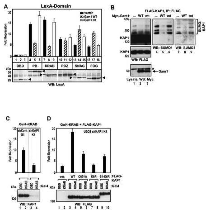
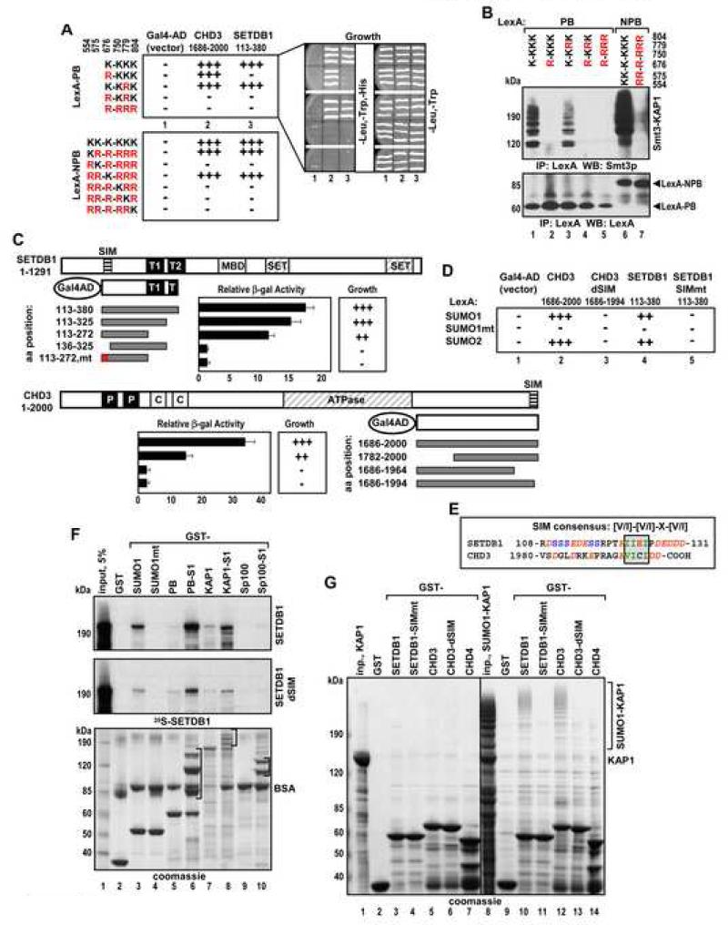
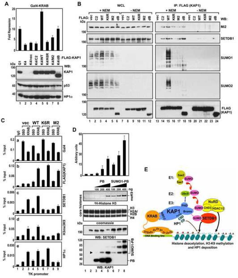
Tandem PHD and bromodomains are often found in chromatin-associated proteins and have been shown to cooperate in gene silencing. Each domain can bind specifically modified histones: the mechanisms of cooperation between these domains are unknown. We show that the PHD domain of the KAP1 corepressor functions as an intramolecular E3 ligase for sumoylation of the adjacent bromodomain. The RING finger-like structure of the PHD domain is required for both Ubc9 binding and sumoylation and directs modification to specific lysine residues in the bromodomain. Sumoylation is required for KAP1-mediated gene silencing and functions by directly recruiting the SETDB1 histone methyltransferase and the CHD3/Mi2 component of the NuRD complex via SUMO-interacting motifs. Sumoylated KAP1 stimulates the histone methyltransferase activity of SETDB1. These data provide a mechanistic explanation for the cooperation of PHD and bromodomains in gene regulation and describe a function of the PHD domain as an intramolecular E3 SUMO ligase.


Figures

Figure 1
Figure 1. KAP1 is Sumoylated in Vivo and in Vitro in a PHD-Dependent Manner
(A) Sequence alignment of six KAP1 sumoylation sites. Forward and reverse sumoylation consensuses are shown and boxed. Major sumoylation lysines are in bold. (B) Schematic representation of KAP1 structural domains. B1 – box 1, B2 – box 2, CC – coiled-coil, HP1BD – HP1 binding domain. Amino acid positions of KAP1 sumoylation site lysines are indicated. Truncations of KAP1 are abbreviated with the first letters of each domain (PB = PHDBromodomain). Sumoylation deficient mutant with all six sumoylation site lysines substituted to arginines is designated RR-R-RRR or K6R. (C) Proteins were immunoprecipitated from H1299 cells with pre-immune serum or antibodies specific for different regions of KAP1: PB or RBCC; or an unrelated LimD1 antibody. A SUMO1 antibody was used in Western blot. Sumoylated forms of KAP1 are indicated with arrows. (D) HEK293 cells were transfected with vector, Ubc9, SENP1 or dnSENP1 plasmids with or without HA-SUMO1 as indicated. Proteins were detected by Western blot with KAP1 antibody. SUMO-KAP1 is indicated with an arrowhead. (E) In vitro sumoylation of GST-KAP1. Immobilized GST and GST-KAP1 were incubated in the presence or absence of recombinant E1, Ubc9, and 32P-SUMO1 as indicated. All in vitro sumoylation reactions described below were analyzed by both Coomassie blue staining (bottom panel) and autoradiography (top panel). (F) HEK293 cells were transfected with the indicated FLAG-KAP1 and T7-SUMO1 plasmids. Proteins were analyzed by Western blot with FLAG antibody. (G-H) In vitro sumoylation of indicated GST-KAP1 proteins. The major mono-sumoylated forms of KAP1 are indicated with arrowheads.
Figure 2
Figure 2. Convergence of Sumoylation Sites and Repression Domains in KAP1
(A) In vitro sumoylation of wild type or sumoylation site KAP1mutants. Arrowheads and arrows indicate the positions of mono- and di-sumoylated forms, respectively. (B-C) HEK293 cells were transfected with plasmids expressing the KAP1 mutants as in (A) fused to LexA, together with a 4xLexA-TK-luc plasmid. LexA-PB plasmids were used in panel C. Data are the mean ± SD of at least two experiments performed in duplicate. (D) HEK293 cells were transfected with T7-SUMO1 and LexA-PB plasmids. LexA-PB proteins were immunoprecipitated with LexA antibody and analyzed by Western blot with SUMO1 (top) and LexA antibodies (bottom). Asterisk – IgGs. (E) HEK293 cells were transfected with the indicated LexA-PB plasmids and wild type or mutant Myc-Gam1 together with 4xLexA-TK-luc plasmid. Protein expression of LexA-PB and Gam1 proteins was confirmed by Western blot (bottom). Data are the mean ± SD of at least two experiments performed in duplicate. (F) In vitro sumoylation of GST-NPB KAP1 proteins. (G) HEK293 cells were transfected with HA-SUMO1 and the indicated LexA-NPB plasmids. Proteins were immunoprecipitated with LexA antibody and analyzed by Western blot using HA (top) and KAP1 antibodies (bottom). (H) HEK293 cells were transfected with the same LexA-NPB plasmids as in (G) with 4xLexATK-luc plasmid. The expression of LexA-NPB proteins was confirmed by Western blot (bottom). Data are the mean ± SD of at least two experiments performed in duplicate.
Figure 3
Figure 3. The KAP1 PHD Binds to Ubc9 with High Selectivity and Directs Sumoylation of the Adjacent Bromodomain
(A) In vitro sumoylation of GST-PB wild type, indicated sumoylation site mutants and C651A mutant proteins. Arrowheads and arrows indicate the positions of mono- and di-sumoylated forms, respectively. (B) HEK293 cells were transfected with indicated LexA-PB plasmids together with 4xLexA-TK-luc plasmid. Data are the mean ± SD of at least two experiments performed in duplicate. (C-D) GST and GST fusions of SUMO1, Ubc9, Sae1 and Sae2 were immobilized on glutathione beads and incubated with recombinant KAP1 PHD. Bound proteins were analyzed by Coomassie blue staining. Lane 1 (C) was loaded with a protein marker in addition to an input sample of the PHD protein. (E-G) In vitro sumoylation of GST- (E) PB modules from KAP1, TIF1α, TIF1γ, Sp100C; (F) RING or RING with adjacent domain (RR) from PIAS1 (aa 140-320), PHD or PB from KAP1; and (G) KAP1 PB and BP chimera.
Figure 4
Figure 4. Mapping of the Ubc9 Binding Site on KAP1 PHD
(A) 2D 1H-15N HSQC spectral comparison of 15N-labeled KAP1 PHD in the free form (black) and in the presence of Ubc9 (red). Molar ratio of KAP1 PHD to Ubc9 is 1:2.4. (Inset) Expansion of spectral region illustrating three peaks that undergo line broadening during Ubc9 titration. The blue peaks correspond to a mid-point of the titration, in which the PHD:Ubc9 molar ratio is 1:1.2. (B) Ribbon and surface representations of the KAP1 PHD structure (PDB code 1FP0), highlighting the residues that exhibited major resonance perturbations upon Ubc9 binding. KAP1 residues corresponding to NMR peaks that undergo line broadening at a PHD:Ubc9 molar ratio of 1:1.2 are in red, and those affected at 1:1.8 are in orange. (C) Sequence alignment of PHD domains from KAP1, TIF1s, BPTF and Sp100C. The KAP1 PHD residues perturbed in the NMR titration experiment are shaded in yellow. Substitutions which inhibit sumoylation (Figure S3) are indicated. (D) Sequence alignment of PHD domains from KAP1 and TIF1γ. The 6 aa substitutions of predicted loop segments L1, L2 and L3 are shown above. Identical residues are shaded in grey. (E) GST, GST-Ubc9 were immobilized on glutathione beads and incubated with the indicated recombinant KAP1 PHDs. Bound proteins were analyzed by Coomassie blue staining. (F) In vitro sumoylation of GST-PB wild type or the indicated loop mutants. (G) U2OS K4 cells were transfected and treated as in Fig.5D. Data are the mean ± SD of at least two experiments performed in duplicate.
Figure 5
Figure 5. Role of KAP1 Sumoylation in KRAB Domain Mediated Repression
(A) HEK293 cells were transfected with the indicated LexA fusions together with vector, wild type or mutant Gam1, and 4xLexA-TK-luc plasmid. The expression of the LexA fusion proteins (arrowheads) was confirmed by Western blot (bottom). (B) HEK293 cells were transfected with FLAG-KAP1 and T7-SUMO1 plasmid together with vector, wild type or mutant Myc-Gam1 plasmids. Gam1 was detected with Myc antibody (bottom). Asterisk - non-specific band. KAP1 was immunoprecipitated with FLAG M2 antibody and Western blotted with FLAG (lanes 1-3), SUMO1 (lanes 4-6) and SUMO3 (lanes 7-9) antibodies. (C) U2OS-G1 and -K4 cells were transfected with Gal4-DBD or Gal4-KRAB plasmids together with 5xGal4-TK-luc plasmid. Endogenous KAP1 protein expression level was verified by Western blot (bottom). (D) U2OS-K4 cells were transfected and treated as in (C) except that vector, FLAG-KAP1 wild type or mutant plasmids were included in the transfection (top). FLAG-KAP1 protein expression was confirmed by Western blot (bottom). (A,C,D) Data are the mean ± SD of at least two experiments performed in duplicate.
Figure 6
Figure 6. SETDB1 and CHD3 Interact with KAP1 in Yeast and in Vitro in SIM Dependent Manner
(A,C,D) Growth selection and quantitative β-galactosidase analyses of L40a yeast cells transformed with the indicated plasmids. The growth was scored on a three-point scale: (+++) - robust growth, (++) - slow growth and (−) – no growth on triple drop-out plates (right). (B) L40a cells transformed with LexA-PB and -NPB plasmids as in (A) were grown in double drop-out media. LexA fusion proteins were immunoprecipitated with LexA antibody and analyzed by Western blot with Smt3p (top) and LexA antibodies (bottom). (C) The domain structure of SETDB1 and CHD3: T1 and T2 – Tudor domains, P – PHD, C – chromodomain. SIM is indicated by a striped box. Data are the mean ± SD from at least three clones analyzed in duplicate. (E) Sequence alignment of SUMO interacting motifs (SIM) from SETDB1 and CHD3. The SIM consensus is boxed and the flanking acidic residues are italicized. (F) Indicated GST fusion proteins immobilized on glutathione beads were incubated with 35S-SETDB1 or SETDB1dSIM. Bound proteins were analyzed by Coomassie blue staining (bottom) and autoradiography (top). Brackets indicate positions of sumoylated isoforms. (G) GST fusions of SETDB1 (aa 113-272) and CHD3 (aa 1782-2000) wild type and SIM mutants, and CHD4 (aa 1768-1912) immobilized on glutathione beads were incubated with recombinant unmodified (lanes 2-7) or sumoylated (lanes 9-14) full length KAP1. Bound proteins were analyzed by Coomassie blue staining. Inp. - input.
Figure 7
Figure 7. KAP1 Sumoylation Promotes Recruitment of SETDB1 to the Target Promoter and Stimulates Its Enzymatic Activity
(A) U2OS-G1, -K4 and indicated reconstituted K4/FLAG-KAP1 cells were treated as in Fig.5C (top). The expression of KAP1, p53 and HP1α proteins was confirmed by Western blot (bottom). Data are the mean ± SD of at least two experiments performed in duplicate. (B) Proteins from reconstituted K4/FLAG-KAP1 cells were immunoprecipitated with FLAG M2 antibodies and Western blotted with Mi2/CHD3, SETDB1, SUMO1, SUMO2 and FLAG antibodies. (C) KAP1-mediated SETDB1 and HP1α recruitment to the TK promoter. U2OS-K4 cells were transfected with Gal4-DBD or Gal4-KRAB, vector, FLAG-KAP1 wild type or indicated mutants together with 5xGal4-TK-luc plasmid and subjected to ChIP analysis. (D) Increasing amounts of unmodified or sumoylated KAP1 PB were mixed with 100 ng of SETDB1 along with 5 μg of core histones and subjected to an in vitro HMTase assay. The arrowhead indicates the position of mono-sumoylated PB. The autoradiograph shows the corresponding 3H-methyl-labeled histone H3. Data are the mean ± SD of three independent experiments (above). (E) A model for sumoylation-dependent, KAP1–mediated gene silencing. The SUMO-conjugated bromodomain recruits the CHD3/NuRD complex and SETDB1 through SUMO-SIM interactions, which results in the deacetylation of histones and the methylation of histone H3-K9. KAP1-bound HP1 recognizes H3-K9 methylation via its chromodomain (CD). Small triangle – acetyl mark, small circle – H3-K9 trimethyl mark on histone tails.

Similar articles

Cited by

References

    1. Boggio R, Colombo R, Hay RT, Draetta GF, Chiocca S. A mechanism for inhibiting the SUMO pathway. Mol Cell. 2004;16:549–561. - PubMed
    1. Bylebyl GR, Belichenko I, Johnson ES. The SUMO isopeptidase Ulp2 prevents accumulation of SUMO chains in yeast. J Biol Chem. 2003;278:44113–44120. - PubMed
    1. Capili AD, Schultz DC, Rauscher IF, Borden KL. Solution structure of the PHD domain from the KAP-1 corepressor: structural determinants for PHD, RING and LIM zinc-binding domains. Embo J. 2001;20:165–177. - PMC - PubMed
    1. Chupreta S, Holmstrom S, Subramanian L, Iniguez-Lluhi JA. A small conserved surface in SUMO is the critical structural determinant of its transcriptional inhibitory properties. Mol Cell Biol. 2005;25:4272–4282. - PMC - PubMed
    1. Friedman JR, Fredericks WJ, Jensen DE, Speicher DW, Huang XP, Neilson EG, Rauscher FJ., 3rd KAP-1, a novel corepressor for the highly conserved KRAB repression domain. Genes Dev. 1996;10:2067–2078. - PubMed

Publication types

MeSH terms

Substances