. 2007 Dec 14;28(5):860-70.
doi: 10.1016/j.molcel.2007.09.021.

Trm9-catalyzed tRNA modifications link translation to the DNA damage response

Affiliations

Trm9-catalyzed tRNA modifications link translation to the DNA damage response

Ulrike Begley et al. Mol Cell. .

Abstract

Transcriptional and posttranslational signals are known mechanisms that promote efficient responses to DNA damage. We have identified Saccharomyces cerevisiae tRNA methyltransferase 9 (Trm9) as an enzyme that prevents cell death via translational enhancement of DNA damage response proteins. Trm9 methylates the uridine wobble base of tRNAARG(UCU) and tRNAGLU(UUC). We used computational and molecular approaches to predict that Trm9 enhances the translation of some transcripts overrepresented with specific arginine and glutamic acid codons. We found that translation elongation factor 3 (YEF3) and the ribonucleotide reductase (RNR1 and RNR3) large subunits are overrepresented with specific arginine and glutamic acid codons, and we demonstrated that Trm9 significantly enhances Yef3, Rnr1, and Rnr3 protein levels. Additionally, we identified 425 genes, which included YEF3, RNR1, and RNR3, with a unique codon usage pattern linked to Trm9. We propose that Trm9-specific tRNA modifications enhance codon-specific translation elongation and promote increased levels of key damage response proteins.


Figures

Figure 1
Figure 1. Trm9 Catalyzed tRNA Modifications
Trm9 completes the formation of mcm5U and mcm5s2U at position 34 of tRNAARG(UCU) and tRNAGLU(UUC), respectively.
Figure 2
Figure 2. Gene-Specific Codon Usage Patterns Identify Codon-Skewed Genes
(A) Hierarchical clustering and heat map analysis of all GSCU data for 5,783 S. cerevisiae genes. Gene-specific over-represented codons are displayed in yellow, under-represented codons displayed in purple, codons with gene-specific codon usage patterns showing no deviation from genome average values displayed as black. Supplemental Figure S3 contains the x-axis key. (B) Northern and Tap tagged western blots for genes and proteins that are over-represented with AGA and GAA codons (i.e., Group 1), and (C) genes outside of Group 1 which contain average codon values. Cells were grown to 5 × 10⁶ cells/ml in YPD and either mock or 0.0125% MMS treated for 1-hour. The loading control (LC) for northern blots was ACT1 while for western blots it is β-tubulin.
Figure 3
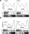
Figure 3. YEF3 and RNR1 transcripts are Associated with Translational Machinery in trm9Δ Cells
(A) Polysome profiles of YEF3 in wild-type and trm9Δ cells. RNA was extracted from the gradient fractions and subjected to northern blot analysis, probing with ACT1 and YEF3 RNA as indicated. (B) Polysome profiles of RNR1 in wild-type and trm9Δ cells. The RNR1 and ACT1 transcripts were detected using northern blot analysis. Nonspecific signal was detected in lanes 10, 13, and 14 and was due to the low RNR1 transcript levels and bleeding from 25S and 18S pools. For all polysome profiles By4741 and trm9Δ cells were grown in YPD to ∼5×10⁶ cells/ml and ethidium bromide staining was used to indicate the relative amounts of 25 and 18S rRNA in each fraction.
Figure 4
Figure 4. Decreased Protein Levels of Rnr1 and Rnr3 Result in a DNA Damage Phenotype
(A) The HU sensitivity of trm9Δ cells was complemented by over-expression of TRM9 from pYES-TRM9. (B) Over-expression of RNR1 and RNR3, from pYES-RNR1 and pYES-RNR3, rescued the MMS sensitive phenotype of trm9Δ cells. For both experiments cell cultures were grown overnight in SD-URA and serially diluted onto plates with HU or MMS, respectively. SD-URA + galactose plates were used for HU assays. YP-galactose plates were used in MMS experiments, because the MMS sensitive phenotype identified in trm9Δ cells is stronger under rich media conditions, compared to defined media.
Figure 5
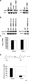
Figure 5. Trm9 Transcript and Protein Levels Are Unchanged after MMS Damage
(A) The transcription of TRM9 in wild-type (BY4741) and trm9Δ cells was analyzed before and after MMS treatment using northern blots. The induction of the MAG1 transcript, which encodes an MMS-inducible DNA glycosylase used to repair 3-methyladenine lesions in DNA, serves as a positive control for DNA damage. (B) Western blots against endogenous TAP tagged Trm9 (ATCC201388 background) before and after MMS treatment. Mag1-TAP (ATCC201388 background) levels increase under these exposure conditions, and serve as a positive control for DNA damage. Cells used for northern and western blots were grown to 5 × 10⁶ cells/ml in YPD and either mock or MMS treated as indicated. (C) Total radioactivity of tRNA purified from wild-type (By4743) and trm9−/− cells labeled with [3H]-methionine before (black bars) and after (white bars) MMS treatment. (D) NaOH derivatization of mcm5U nucleosides will generate labeled methanol. NaOH reactive methylesters in tRNA from wild-type (BY4743) and trm9−/− cells labeled with [3H]-methionine, before (black bars) and after (white bars) MMS treatment. Error bars represent the standard deviation of two replicate experiments in panels C and D.
Figure 6
Figure 6. Model for Codon-Specific Translational Enhancement
Our model has (panel A, left) Trm9 dependent tRNA modifications enhancing the translation of transcripts over-represented with AGA codons while not affecting average transcripts. Translational enhancement is most likely facilitated (panel A, right) by mcm5U (*) at the tRNA wobble base promoting interactions with the AGA in mRNA. It is important to note that the up- and down-stream codons surrounding AGA most likely play an important yet still undefined role in this model. The lack of Trm9-dependent tRNA modifications (panel B, left) slows the translation of transcripts over-represented with AGA codons. Codon-specific translation is predicted to be slowed due to (panel B, right) inefficient interactions between unmodified uridine in the anticodon and adenine in the codon wobble position. AGA specific transcripts and proteins are shown in yellow, while average transcripts and proteins are shown in gray.

Comment in

  • Better living through biochemistry.
    Eisenstein M. Eisenstein M. Nat Methods. 2011 Feb;8(2):108-9. doi: 10.1038/nmeth0211-108b. Nat Methods. 2011. PMID: 21355118 No abstract available.

Similar articles

Cited by

References

    1. Agris PF. Decoding the genome: a modified view. Nucleic Acids Res. 2004;32:223–238. - PMC - PubMed
    1. Alexandrov A, Grayhack EJ, Phizicky EM. tRNA m7G methyltransferase Trm8p/Trm82p: Evidence linking activity to a growth phenotype and implicating Trm82p in maintaining levels of active Trm8p. RNA. 2005;11:821–30. - PMC - PubMed
    1. Alexandrov A, Chernyakov I, Gu W, Hiley SL, Hughes TR, Grayhack EJ, Phizicky EM. Rapid tRNA decay can result from lack of nonessential modifications. Mol Cell. 2006;21:87–96. - PubMed
    1. Anand M, Chakraburtty K, Marton MJ, Hinnebusch AG, Kinzy TG. Functional interactions between yeast translation eukaryotic elongation factor (eEF) 1A and eEF3. J Biol Chem. 2003;278:6985–6991. - PubMed
    1. Barrows LR, Magee PN. Nonenzymatic methylation of DNA by S-adenosylmethionine in vitro. Carcinogenesis. 1982;3:349–351. - PubMed

Publication types

MeSH terms