. 2008 Mar 3;152(1):273-87.
doi: 10.1016/j.neuroscience.2007.10.042.

Lipopolysaccharide suppresses activation of the tuberomammillary histaminergic system concomitant with behavior: a novel target of immune-sensory pathways

Affiliations

Lipopolysaccharide suppresses activation of the tuberomammillary histaminergic system concomitant with behavior: a novel target of immune-sensory pathways

R P A Gaykema et al. Neuroscience. .

Abstract

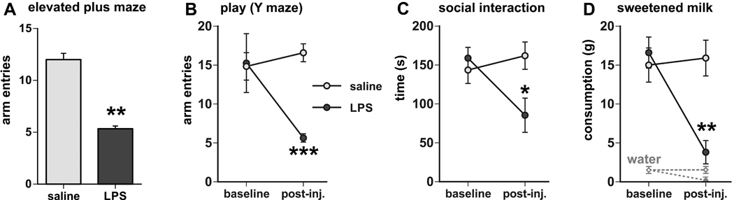
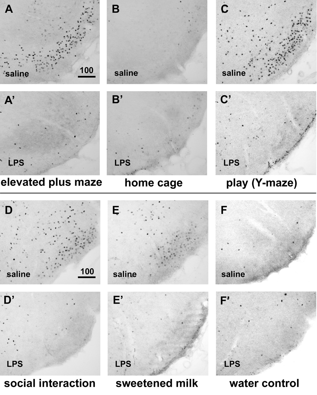
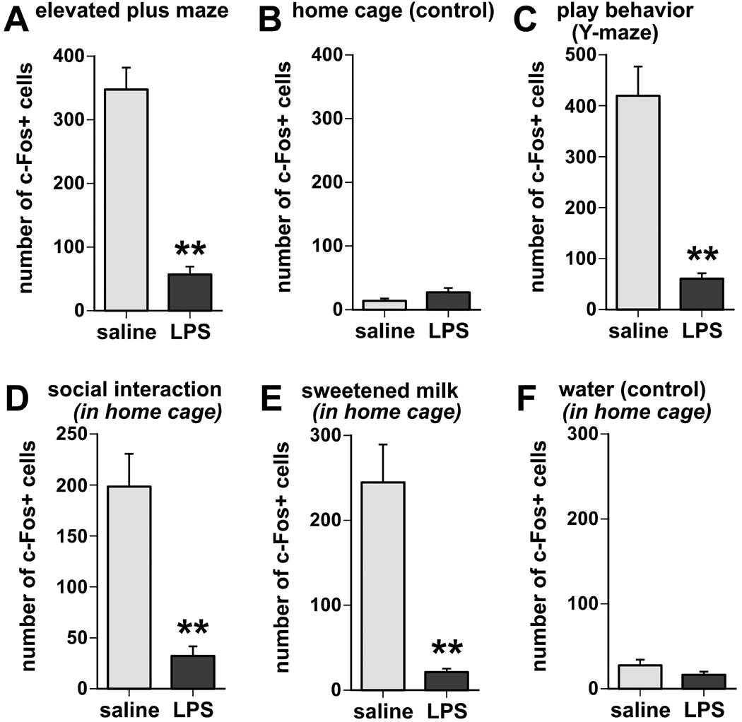
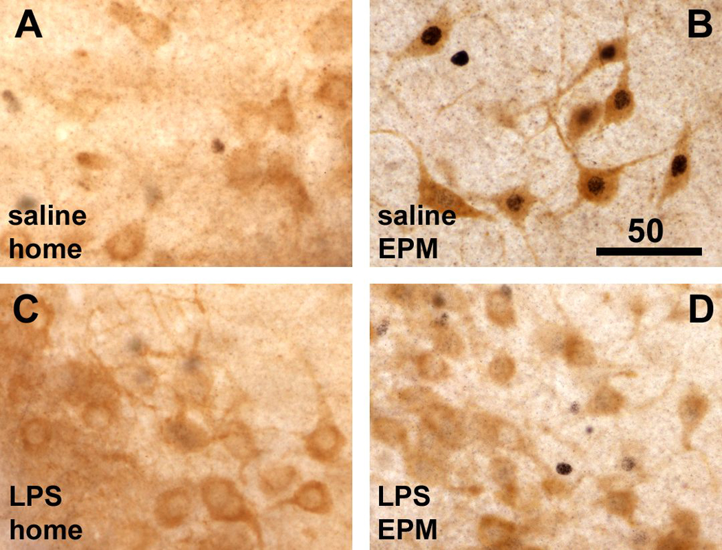
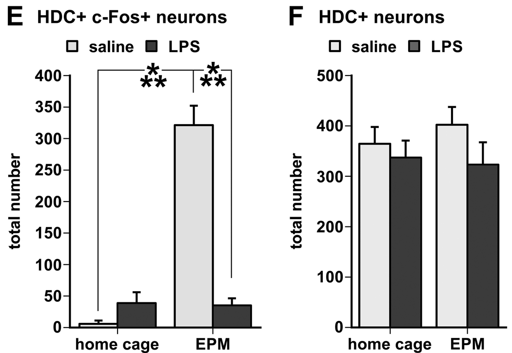
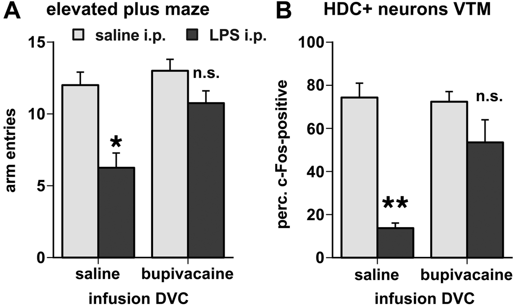
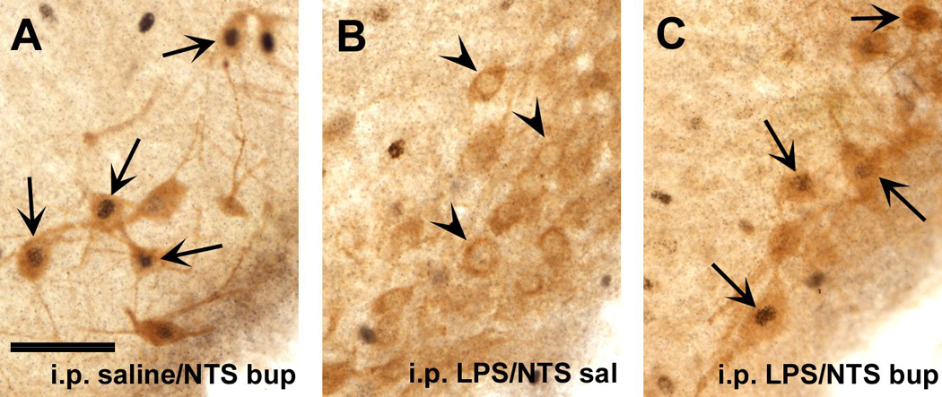
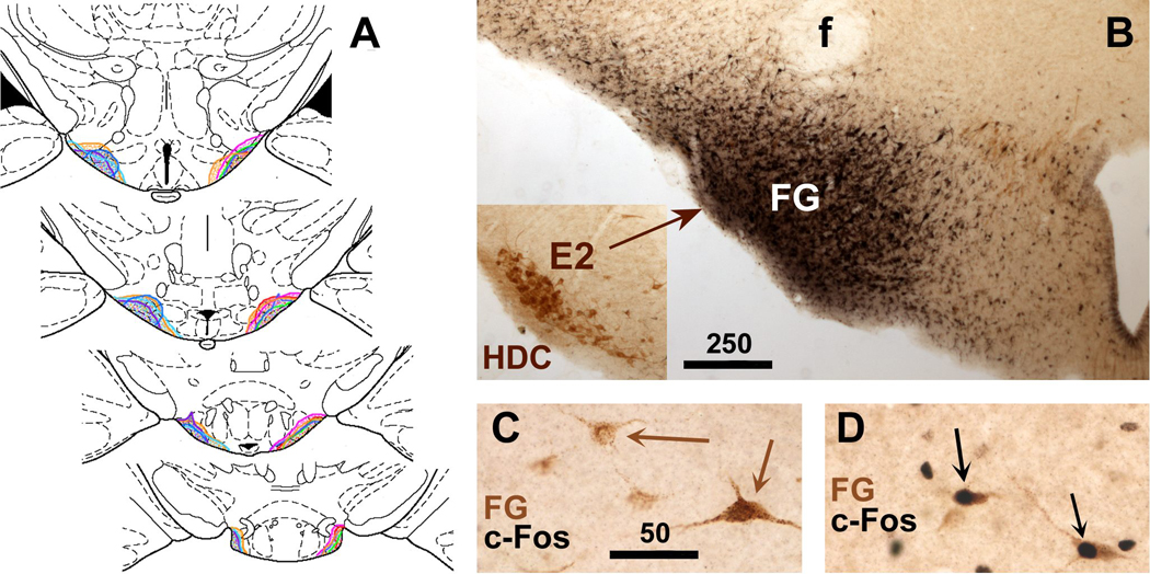
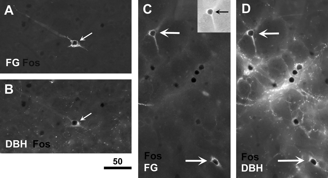
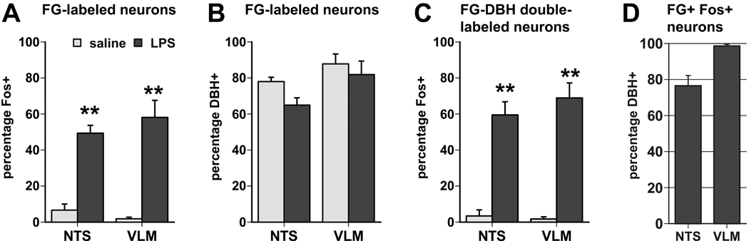
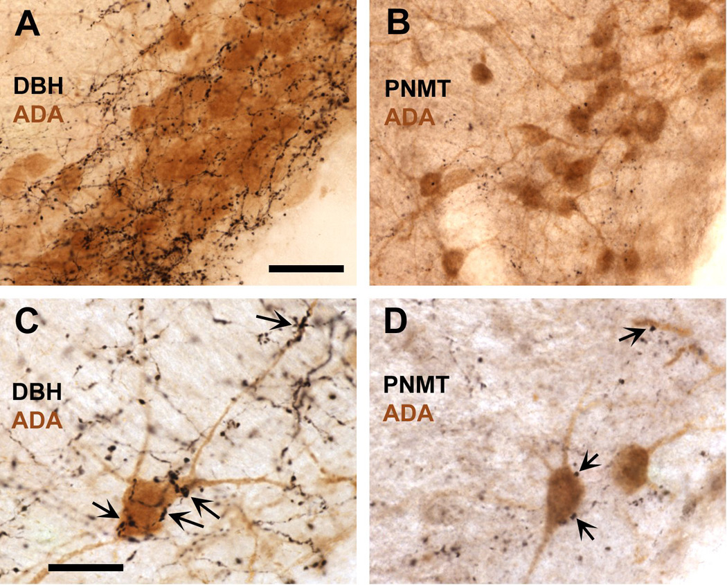
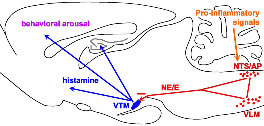
Infection and inflammation strongly inhibit a variety of behaviors, including exploration, social interaction, and food intake. The mechanisms that underlie sickness behavior remain elusive, but appear to involve fatigue and a state of hypo-arousal. Because histaminergic neurons in the ventral tuberomammillary nucleus of the hypothalamus (VTM) play a crucial role in the mediation of alertness and behavioral arousal, we investigated whether the histaminergic system represents a target for immune activation and, if so, whether modulation by ascending medullary immune-sensitive projections represents a possible mechanism. Rats were injected intraperitoneally with either the pro-inflammatory stimulus lipopolysaccharide (LPS) or saline, and exposed to one of various behavioral tests that would induce motivated behavior (exploration, play behavior, social interaction, sweetened milk consumption). Upon kill, brains were processed for c-Fos and histidine decarboxylase immunoreactivity. LPS treatment reduced behavioral activity and blocked behavioral test-associated c-Fos induction in histaminergic neurons of the VTM. These effects of LPS were prevented by prior inactivation of the caudal medullary dorsal vagal complex (DVC) with a local anesthetic. To determine whether LPS-responsive brainstem projection neurons might provide a link from the DVC to the VTM, the tracer Fluorogold was iontophoresed into the VTM a week prior to experiment. Retrogradely labeled neurons that expressed c-Fos in response to LPS treatment included catecholaminergic neurons within the nucleus of the solitary tract and ventrolateral medulla. These findings support the hypothesis that the histaminergic system represents an important component in the neurocircuitry relevant for sickness behavior that is linked to ascending pathways originating in the lower brainstem.


Figures

Fig. 1
Fig. 1
Behavioral activity is suppressed by prior treatment with LPS in the elevated plus maze-novel environment exploration task, shown by the reduced number of arm entries (A). Similarly, LPS treatment suppressed behavioral activity during playful activity in the Y maze (reduced number of arm entries in B), during social interaction with a conspecific juvenile (reduced interaction time in C), and during the presentation of sweetened milk (reduced consumption in D). The graphs in B-D display the baseline values of the behavioral measures (averaged over the two days prior to the day of experiment) in addition to the values following the injections of saline or LPS (post-inj.). * p < 0.05, ** p < 0.005, *** p < 0.0005, LPS vs. saline treatment.
Fig. 2
Fig. 2
Induction of c-Fos immunoreactivity in the VTM associated with behavioral activation and LPS-induced suppression. In saline-treated subjects, both exploration of a novel environment (elevated plus maze, in A) and playful behavior (Y-maze, in C) induced strong neuronal c-Fos expression that was absent in control animals left in the home cage (B). Behavioral activity in the home cage such as social interaction (D) and consumption of sweetened milk (E) also lead to induction, albeit more modest, of c-Fos immunoreactivity in the VTM, which was absent in rats offered water instead (F). After LPS treatment, little c-Fos induction could be discerned in the VTM irrespective of the behavioral context the animals were exposed to (EPM: A’; play/Y-maze: C’; social interaction: D’; sweetened milk: E’), and the staining could not be distinguished from that of LPS-treated rats left undisturbed in the home cage (B’) or those offered water (F’). Scale bar in micrometers.
Fig. 3
Fig. 3
Semi-quantitative analysis of c-Fos-positive nuclei in the VTM reveals that LPS treatment strongly inhibited the induction of c-Fos immunoreactivity associated with exploratory (A), play behavior (B), social interaction (D), and sweetened milk consumption (D), whereas there was little c-Fos induction, and little or no difference between LPS and saline treatments, in home cage controls (B) or when offered water instead of sweetened milk (F). Values expressed represent means and SEM of cumulative counts in 4 sections through the posterior hypothalamus containing the E1-E3 regions of the VTM. ** p < 0.005, LPS vs.saline treatment.
Fig. 4
Fig. 4
Double labeling for cytoplasmic L-histidine decarboxylase (HDC) and nuclear c-Fos immunoreactivity shows that HDC-positive neurons lacked nuclear c-Fos staining in home cage animals (A), but displayed strong nuclear staining for c-Fos (black) in rats 1 hour following EPM exploration (B). LPS challenge itself had little effect on c-Fos expression in HDC-positive neurons in home cage controls (C), but prior to the EPM test LPS challenge almost completely inhibited nuclear staining for c-Fos in HDC-positive neurons despite behavioral activity exhibited by the LPS-treated rats (D). Scale bar in B in µm. E. Semi-quantitative assessment of c-Fos immunoreactivity in HDC-positive (histaminergic) neurons revealed a strong induction in rats subjected to the exploration task, which was reversed by LPS challenge 90 minutes prior to the test (***, p < 0.0001). F. There was no difference between groups in the total number of HDC-positive cell bodies with visible cell nuclei (either c-Fos-labeled or unlabeled).
Fig. 4
Fig. 4
Double labeling for cytoplasmic L-histidine decarboxylase (HDC) and nuclear c-Fos immunoreactivity shows that HDC-positive neurons lacked nuclear c-Fos staining in home cage animals (A), but displayed strong nuclear staining for c-Fos (black) in rats 1 hour following EPM exploration (B). LPS challenge itself had little effect on c-Fos expression in HDC-positive neurons in home cage controls (C), but prior to the EPM test LPS challenge almost completely inhibited nuclear staining for c-Fos in HDC-positive neurons despite behavioral activity exhibited by the LPS-treated rats (D). Scale bar in B in µm. E. Semi-quantitative assessment of c-Fos immunoreactivity in HDC-positive (histaminergic) neurons revealed a strong induction in rats subjected to the exploration task, which was reversed by LPS challenge 90 minutes prior to the test (***, p < 0.0001). F. There was no difference between groups in the total number of HDC-positive cell bodies with visible cell nuclei (either c-Fos-labeled or unlabeled).
Fig. 5
Fig. 5
A. Inactivation of the dorsal vagal complex reversed the inhibitory effects of LPS challenge on behavioral activity during exploration of the elevated plus maze, as measured by the total number of arm entries. B. Inactivation of the DVC also diminished the suppressive effect of LPS on the c-Fos induction in histaminergic neurons associated with the exploratory behavior (LPS effects: *, p < 0.01; **, p < 0.001; n.s., not significant).
Fig. 6
Fig. 6
A. Induction of c-Fos immunoreactivity (black nuclear staining) in HDC-immunoreactive neurons in the VTM (lighter brown cytoplasmic staining) of a saline-treated rat following exploration of the elevated plus maze. Arrows point at double-labeled neurons. B,C. Following i.p. LPS challenge and maze exposure, c-Fos staining was absent in most HDC-labeled neurons, when saline was infused into the DVC (arrowheads in B), but appeared in HDC-positive neurons in LPS-treated rat that received intra-DVC bupivacaine infusion (arrows in C).
Fig. 7
Fig. 7
A. The extent of FG injection sites in the VTM shown in brain section diagrams (Paxinos and Watson, 1998) at 4 rostrocaudal levels through the posterior hypothalamus. FG injections of individual cases are indicated by color, and assembled together to show the rostrocaudal extent in each case. B. Photomicrograph of a representative FG injection site aimed at the VTM. FG immunoreactivity was visualized with black staining using nickelous DAB, whereas HDC immunoreactivity was revealed using regular DAB, yielding brown staining. The inserted photomicrograph shows HDC-containing neurons in the same location, taken from the opposite hemisphere in the same section, and presented after being flipped 180 degrees. C,D: In the NTS, retrogradely labeled neurons (displaying FG immunoreactivity stained brown using DAB) show black c-Fos immunostaining only after intraperitoneal LPS challenge (black arrows in D), but not after saline injection (brown arrows in C). Scale bars in B,C in micrometers.
Fig. 8
Fig. 8
FG-positive neurons (retrogradely labeled from the VTM region) in the NTS (A,B) and VLM (C,D) that display LPS-induced c-Fos expression are predominantly DBH-positive. The triple staining was achieved by sequential immunofluoresence labeling for FG (Alexa Fluor 488 excitation, shown in A,C) and DBH (Alexa Fluor 546 excitation, shown in B,D), followed by initial immunoperoxidase staining for c-Fos as revealed by the intense black shadow (arrows) or staining visualized by complementary brightfield illumination (insert in the top right corner of panel C). The micrographs in A and B, and those in C and D, captured the same location in the NTS and VLM, respectively. Scale bar in micrometers.
Fig. 9
Fig. 9
A. Semi-quantitative analysis of the induction of c-Fos expression by LPS challenge (0.1 mg/kg i.p.), but not saline injection, in FG-labeled neurons in the NTS and VLM (retrogradely labeled from the VTM region of the posterior hypothalamus). Values are expressed as the fraction of the total of retrogradely labeled neurons with a prominently visible nucleus in the plane of the section (**, p < 0.005). B. The FG-labeled neurons in the NTS and VLM predominantly show a noradrenergic/adrenergic phenotype as most display DBH immunoreactivity. C. LPS-mediated induction of c-Fos expression occurs in 60–70% of all FG-DBH-double-labeled neurons encountered in the NTS and VLM. D. Of all the neurons double-labeled for FG and c-Fos following LPS treatment, the majority in the NTS (77%) and virtually all in the VLM (99%) are also labeled for DBH immunoreactivity.
Fig. 10
Fig. 10
Rich catecholaminergic innervation in the histaminergic cell-rich ventral tuberomammillary nucleus as demonstrated by the dense plexus of DBH-immunoreactive varicose fibers (black) intermingling with adenosine deaminase (ADA)-positive cells (brown), a marker for histaminergic neurons in the VTM (A). Although considerably less dense, the VTM is also supplied by rather fine PNMT-positive fibers, vaticosities and terminal boutons (B), indicative of adrenergic innervation, which is exclusively derived from the medulla. Both the DBH- and PNMT-labeled fibers and varicosities are predominantly distributed amongst the ADA-positive cells in the VTM, and are much sparser in the adjacent portions of the ventral posterior hypothalamus. Photomicrographs taken under higher magnification reveal close appositions of DBH- and PNMT- labeled varicosities (C and D, respectively) with ADA-immunreactive cell bodies and dendrites, indicative of a direct catecholaminergic influence on these cells. Scale bar in A = 50 µm, also applies to B; scale bar in C = 25 µm, also applies to D.
Fig. 11
Fig. 11
Summary model based on the present findings. The brain diagram depicting the inhibitory, primarily catecholaminergic influence, arising from the NTS and VLM, on the histaminergic system in the ventral tuberomammillary nucleus (VTM) of the hypothalamus. This inhibitory influence is initiated by a peripheral inflammatory stimulus (like LPS), and result in diminished behavioral arousal, a process highly dependent on activation of the histaminergic system.

Similar articles

Cited by

References

    1. Aiche SA, Saravay RH, Cravo S, Jeske I, Morrison SF, Reis DJ, Milner TA. Monosynaptic projections from the nucleus tractus solitarii to C1 adrenergic neurons in the rostral ventrolateral medulla: comparison with input from the caudal ventrolateral medulla. J Com Neurol. 1996;373:62–75. - PubMed
    1. Becskei C, Riediger T, Hernadfalvy N, Arsenijevic D, Lutz TA, Langhans W. Inhibitory effects of lipopolysaccharide on hypothalamic nuclei implicated in the control of food intake. Brain Behav Immun. 2007 epub ahead of print. - PubMed
    1. Buller K, Xu Y, Dayas C, Day T. Dorsal and ventral medullary catecholamine cell groups contribute differentially to systemic interleukin-1β-induced hypothalamic pituitary adrenal axis responses. Neuroendocrinol. 2001;73:129–138. - PubMed
    1. Buller KM, Day TA. Systemic administration of interleukin-1β activates select populations of central amygdala afferents. J Comp Neurol. 2002;452:288–296. - PubMed
    1. Burgdorf J, Panksepp J. Tickling induces reward in adolescent rats. Physiol Behav. 2001;72:167–173. - PubMed

Publication types