

Activity-dependent secretion of neuronal activity regulated pentraxin from vasopressin neurons into the systemic circulation
- PMID: 18082971
- PMCID: PMC2342909
- DOI: 10.1016/j.neuroscience.2007.10.040
Activity-dependent secretion of neuronal activity regulated pentraxin from vasopressin neurons into the systemic circulation
Abstract
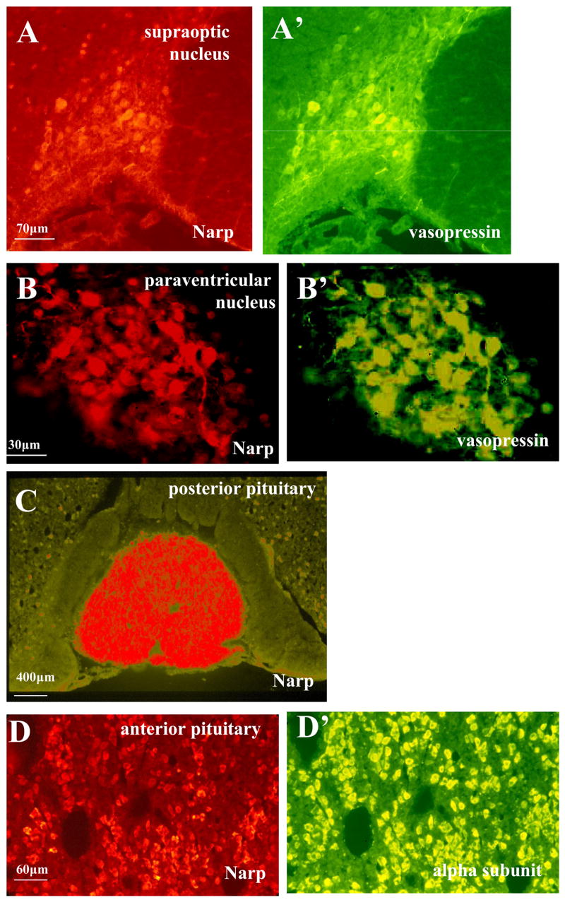
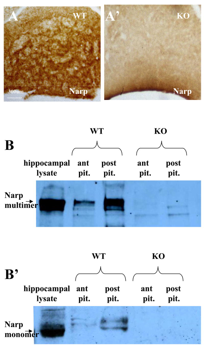
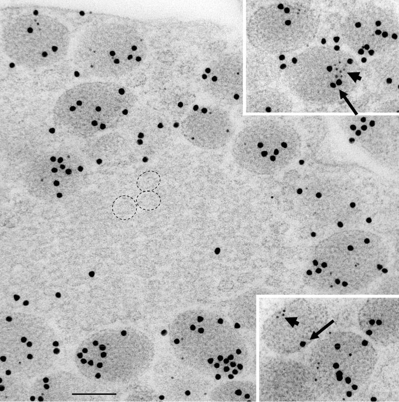
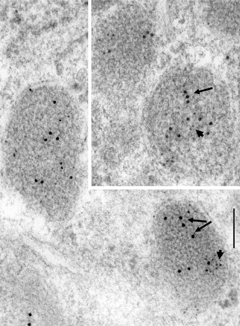
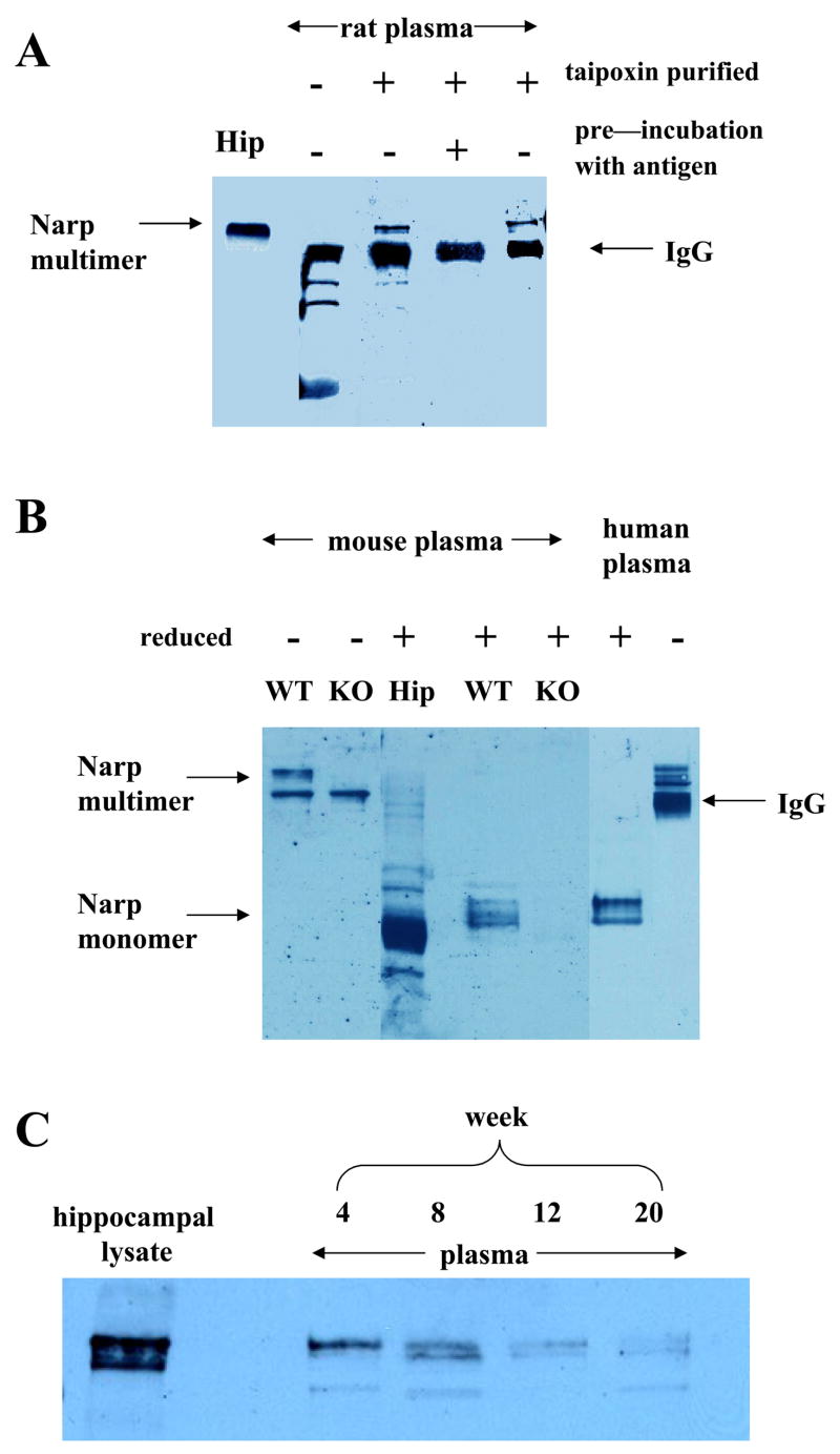
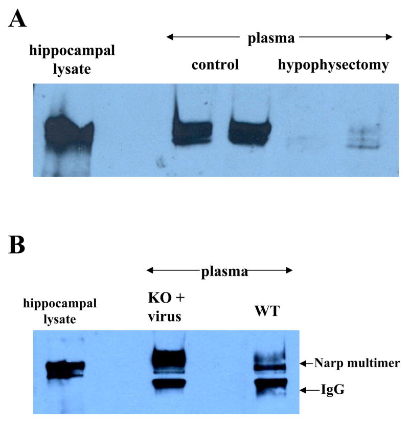
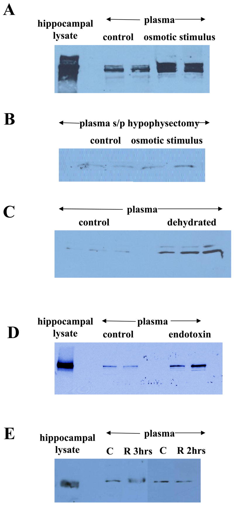
Neuronal activity regulated pentraxin (Narp) is a secreted, synaptic protein that has been implicated in modulating synaptic transmission. However, it is unclear how Narp secretion is regulated. Since we noted prominent Narp immunostaining in vasopressin neurons of the hypothalamus and in the posterior pituitary, we assessed whether it, like vasopressin, is released into the systemic circulation in an activity-dependent fashion. Consistent with this hypothesis, electron microscopic studies of the posterior pituitary demonstrated that Narp is located in secretory vesicles containing vasopressin. Using affinity chromatography, we detected Narp in plasma and found that these levels are markedly decreased by hypophysectomy. In addition, we confirmed that injection of a viral Narp construct into the hypothalamus restores plasma Narp levels in Narp knockout mice. In checking for activity-dependent secretion of Narp from the posterior pituitary, we found that several stimuli known to trigger vasopressin release, i.e. hypovolemia, dehydration and endotoxin, elevate plasma Narp levels. Taken together, these findings provide compelling evidence that Narp is secreted from vasopressin neurons in an activity-dependent fashion.
Figures
Similar articles
-
Localized disruption of Narp in medial prefrontal cortex blocks reinforcer devaluation performance.Learn Mem. 2010 Nov 22;17(12):620-6. doi: 10.1101/lm.1937210. Print 2010 Dec. Learn Mem. 2010. PMID: 21127001 Free PMC article.
-
Selective expression of Narp, a secreted neuronal pentraxin, in orexin neurons.J Neurochem. 2002 Sep;82(6):1561-5. doi: 10.1046/j.1471-4159.2002.01141.x. J Neurochem. 2002. PMID: 12354306
-
Neuronal pentraxins modulate cocaine-induced neuroadaptations.J Pharmacol Exp Ther. 2009 Jan;328(1):183-92. doi: 10.1124/jpet.108.143115. Epub 2008 Oct 7. J Pharmacol Exp Ther. 2009. PMID: 18840757 Free PMC article.
-
State-dependent plasticity in vasopressin neurones: dehydration-induced changes in activity patterning.J Neuroendocrinol. 2010 May;22(5):343-54. doi: 10.1111/j.1365-2826.2010.01961.x. Epub 2010 Jan 19. J Neuroendocrinol. 2010. PMID: 20088912 Review.
-
[Role of the neurohypophysis in psychological stress].Encephale. 2001 May-Jun;27(3):245-59. Encephale. 2001. PMID: 11488255 Review. French.
Cited by
-
Activity-dependent diffusion trapping of AMPA receptors as a key step for expression of early LTP.Philos Trans R Soc Lond B Biol Sci. 2024 Jul 29;379(1906):20230220. doi: 10.1098/rstb.2023.0220. Epub 2024 Jun 10. Philos Trans R Soc Lond B Biol Sci. 2024. PMID: 38853553 Free PMC article. Review.
-
Foxa1 and Foxa2 maintain the metabolic and secretory features of the mature beta-cell.Mol Endocrinol. 2010 Aug;24(8):1594-604. doi: 10.1210/me.2009-0513. Epub 2010 Jun 9. Mol Endocrinol. 2010. PMID: 20534694 Free PMC article.
-
Selective expression of Narp in primary nociceptive neurons: role in microglia/macrophage activation following nerve injury.J Neuroimmunol. 2014 Sep 15;274(1-2):86-95. doi: 10.1016/j.jneuroim.2014.06.016. Epub 2014 Jun 28. J Neuroimmunol. 2014. PMID: 25005116 Free PMC article.
-
Narp regulates long-term aversive effects of morphine withdrawal.Behav Neurosci. 2008 Aug;122(4):760-8. doi: 10.1037/a0012514. Behav Neurosci. 2008. PMID: 18729628 Free PMC article.
-
Regulation of AMPA receptor trafficking by secreted protein factors.Front Cell Neurosci. 2023 Nov 27;17:1271169. doi: 10.3389/fncel.2023.1271169. eCollection 2023. Front Cell Neurosci. 2023. PMID: 38089145 Free PMC article. Review.
References
-
- Boldyrev AA, Carpenter DO, Johnson P. Emerging evidence for a similar role of glutamate receptors in the nervous and immune systems. J Neurochem. 2005;95:913–8. - PubMed
-
- de Bree FM. Trafficking of the vasopressin and oxytocin prohormone through the regulated secretory pathway. J Neuroendocrinol. 2000;12:589–94. - PubMed
-
- Burger C, Gorbatyuk OS, Velardo MJ, Peden CS, Williams P, Zolotukhin S, Reier PJ, Mandel RJ, Muzyczka N. Recombinant AAV viral vectors pseudotyped with viral capsids from serotypes 1, 2, and 5 display differential efficiency and cell tropism after delivery to different regions of the central nervous system. Mol Ther. 2004;10:302–17. - PubMed
Publication types
MeSH terms
Substances
Grants and funding
LinkOut - more resources
Full Text Sources
Molecular Biology Databases
