. 2008 Mar 30;373(1):189-201.
doi: 10.1016/j.virol.2007.11.001. Epub 2008 Feb 20.

Interactions of LSECtin and DC-SIGN/DC-SIGNR with viral ligands: Differential pH dependence, internalization and virion binding

Affiliations

Interactions of LSECtin and DC-SIGN/DC-SIGNR with viral ligands: Differential pH dependence, internalization and virion binding

Thomas Gramberg et al. Virology. .

Abstract

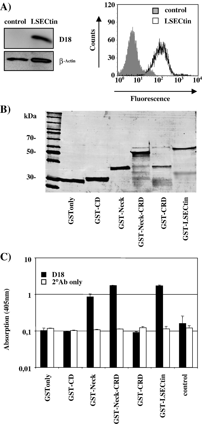
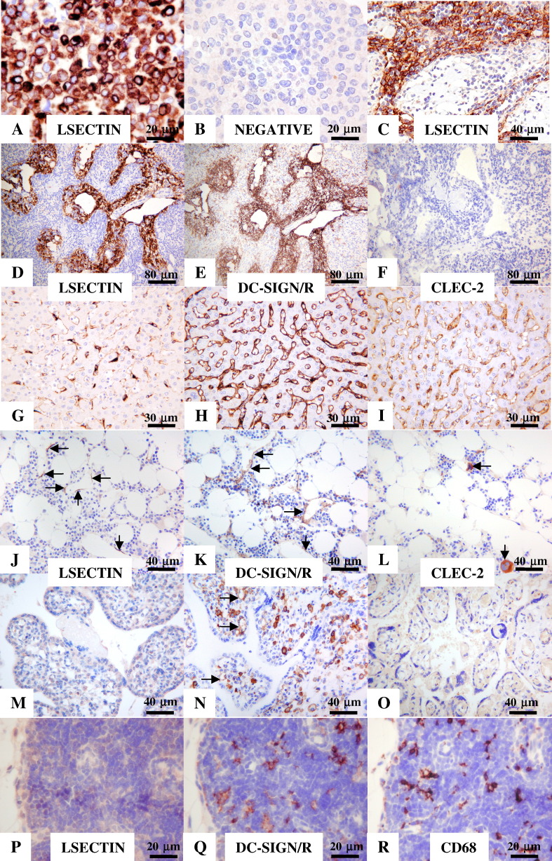
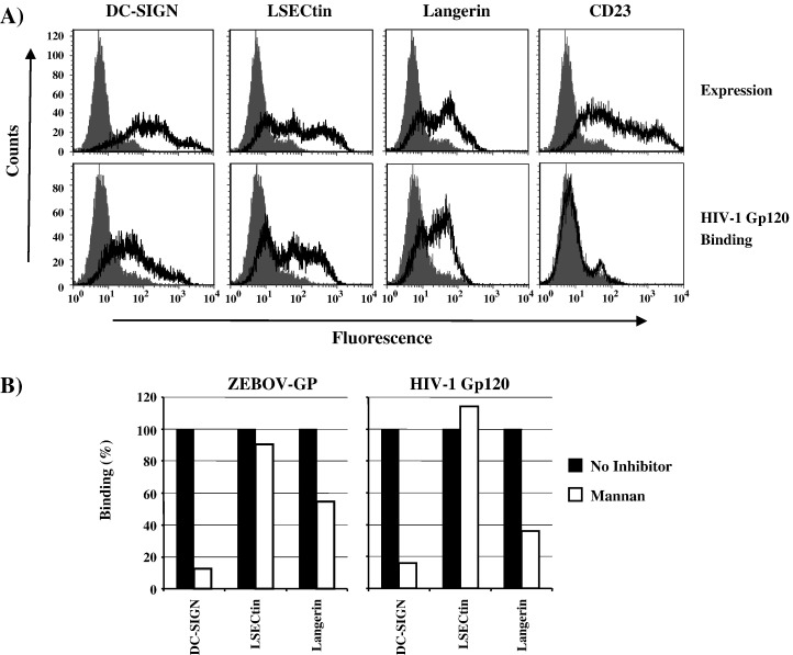
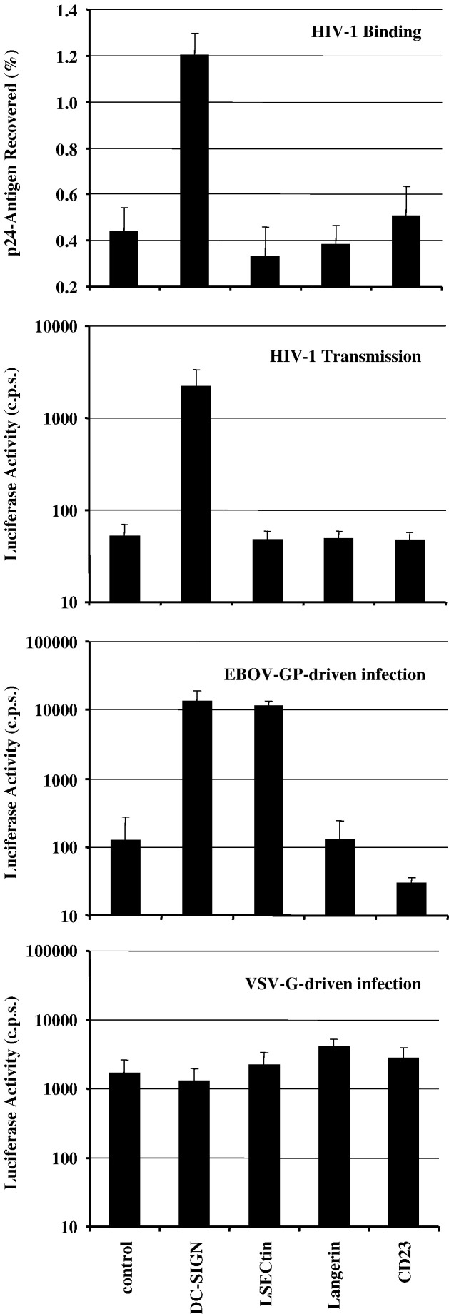
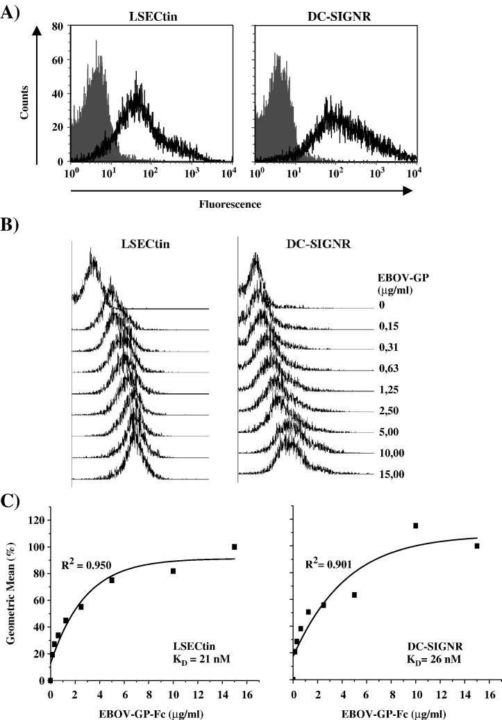
The calcium-dependent lectins DC-SIGN and DC-SIGNR (collectively termed DC-SIGN/R) bind to high-mannose carbohydrates on a variety of viruses. In contrast, the related lectin LSECtin does not recognize mannose-rich glycans and interacts with a more restricted spectrum of viruses. Here, we analyzed whether these lectins differ in their mode of ligand engagement. LSECtin and DC-SIGNR, which we found to be co-expressed by liver, lymph node and bone marrow sinusoidal endothelial cells, bound to soluble Ebola virus glycoprotein (EBOV-GP) with comparable affinities. Similarly, LSECtin, DC-SIGN and the Langerhans cell-specific lectin Langerin readily bound to soluble human immunodeficiency virus type-1 (HIV-1) GP. However, only DC-SIGN captured HIV-1 particles, indicating that binding to soluble GP is not necessarily predictive of binding to virion-associated GP. Capture of EBOV-GP by LSECtin triggered ligand internalization, suggesting that LSECtin like DC-SIGN might function as an antigen uptake receptor. However, the intracellular fate of lectin-ligand complexes might differ. Thus, exposure to low-pH medium, which mimics the acidic luminal environment in endosomes/lysosomes, released ligand bound to DC-SIGN/R but had no effect on LSECtin interactions with ligand. Our results reveal important differences between pathogen capture by DC-SIGN/R and LSECtin and hint towards different biological functions of these lectins.


Figures

Fig. 1
Fig. 1
Characterization of the LSECtin-specific monoclonal antibody D18. (A) D18 allows detection of LSECtin by Western blot and FACS analysis. LSECtin was transiently expressed in 293T cells and expression detected by Western blot (left panel) or FACS (right panel). Cells transfected with empty vector were analyzed as negative control. Staining for β-actin served as loading control for the Western blot analysis. (B) Analysis of recombinant LSECtin variants. Wild-type LSECtin or the indicated LSECtin domains fused to GST were purified from bacteria, separated by SDS–gel electrophoresis and visualized by Coomassie staining. (C) The D18 epitope is located in the LSECtin neck region. The indicated recombinant LSECtin proteins were coated onto 96-well plates and D18 reactivity against the proteins was analyzed by ELISA. D18 staining of uncoated control wells and staining with secondary antibody alone served as negative controls. The results of a representative experiment are shown and were confirmed in two separate experiments.
Fig. 2
Fig. 2
LSECtin is co-expressed with DC-SIGNR on liver, lymph node and bone marrow sinusoidal endothelial cells. Expression of LSECtin on stably transfected 293T-REx cells was induced by doxycycline (A) or PBS (B) and the cells were subsequently formalin fixed, paraffin embedded and immunostained (brown) for LSECtin. Nuclei were stained with hematoxylin (blue). Lymph node was immunostained (brown) for LSECtin (C and D), DC-SIGN/R (E) and CLEC-2 (F). Liver sections immunostained for LSECtin (G), DC-SIGN/R (H) and CLEC-2 (I) demonstrated a continuous staining pattern around hepatic sinusoids for all the lectin molecules, indicative of expression of all three molecules by sinusoidal endothelial cells. Serial sections of bone marrow demonstrated expression of LSECtin (arrows on J) and DC-SIGN/R (arrows on K), but not CLEC-2 (L) on sinusoidal endothelial cells. DC-SIGN was also expressed by some megakaryocytes (K) and possibly other bone marrow cell types, whereas CLEC-2 was expressed by megakaryocytes only (arrows on L). Sections of placenta were negative for LSECtin (M) and CLEC-2 (O) but demonstrated DC-SIGN/R expression by vascular endothelial cells (arrows on N) and DC-SIGN expression by Hofbauer cells (N). Sections of thymus showed no expression of LSECtin (P), although abundant macrophages/dendritic cells were demonstrated by immunostaining for DC-SIGN/R (Q) and CD68 (R).
Fig. 3
Fig. 3
LSECtin binds to soluble HIV-1-Gp120 in a mannose independent fashion. (A) HeLa cells were transiently transfected with plasmids encoding the indicated lectins and lectin expression and HIV-1-Gp120 binding analyzed in parallel. Lectin expression was determined by staining with an antibody directed against a C-terminal AU1 antigenic tag (DC-SIGN, Langerin, CD23) or with the LSECtin-specific monoclonal antibody D18 (top panel). Lectin binding to an HIV-1-Gp120-Fc fusion protein was assessed by incubating lectin expressing cells with concentrated supernatant containing HIV-1-Gp120 followed by detection of bound protein with an Fc-specific antibody (bottom panel). Similar results were obtained in two separate experiments. (B) Inhibition of HIV-1-Gp120 binding by mannan. Binding of HIV-1-Gp120 to lectin expressing cells was analyzed as described for panel (A); however, cells were pre-incubated with PBS or mannan before addition of soluble envelope protein. Binding to lectin expressing cells in the absence of inhibitor was set as 100%. The results of a representative experiment are shown and were confirmed in two independent experiments.
Fig. 4
Fig. 4
LSECtin does not capture and transmit HIV-1 to target cells. HeLa cells transiently expressing the indicated lectins or control transfected cells were pulsed with HIV-1 or HIV-1 pseudotypes bearing EBOV-GP or VSV-G. All viruses encoded the luciferase gene in place of nef. In order to determine HIV-1 binding, the cells were washed and the amount of p24-antigen in cell lysates determined (top panel). Results are shown as percent of recovered input antigen. HIV-1 transmission was assessed by coculture of virus-exposed cells with target cells followed by measurement of luciferase activities in culture lysates (second panel). The efficiency of EBOV-GP and VSV-G-driven infection was determined by quantifying luciferase activities in the lysates of infected HeLa cells (third and fourth panel). The results of representative experiments carried out in triplicate are presented. Error bars indicate standard deviation (SD). Similar results were obtained in two independent experiments.
Fig. 5
Fig. 5
LSECtin and DC-SIGNR exhibit similar affinities for EBOV-GP. 293T-REx cells (Pöhlmann et al., 2001a, Pöhlmann et al., 2001b, Simmons et al., 2003) were induced to express comparable amounts of LSECtin or DC-SIGNR, incubated with the indicated amounts of purified soluble EBOV-GP-Fc fusion protein and binding determined by FACS. Results of a representative experiment are shown in (A, lectin expression) and (B, GP binding). The average of four independent experiments, for which the relative geometric mean channel fluorescence values were determined, is shown in panel C. Geometric mean values observed under saturating conditions were set as 100%. Error bars indicate standard deviation. The results were used to calculate KD values employing a previously described method (Lozach et al., 2003).
Fig. 6
Fig. 6
LSECtin internalizes EBOV-GP and lectin–ligand complexes are not dissolved by low pH. (A) LSECtin internalizes ligand. B-THP cells engineered to express DC-SIGN or LSECtin or control B-THP cells (Wu et al., 2004) were incubated with Alexa647-labeled soluble EBOV-GP-Fc fusion protein on ice; subsequently, cells were kept on ice or shifted to 37 °C for 5 min or 15 min to allow internalization of bound antigen. After trypsin or control treatment lectin expression and EBOV-GP binding were analyzed. The results of a representative experiment are shown and were confirmed in two separate experiments. (B) Lysosomal pH does not dissociate LSECtin–ligand complexes. 293T-REx cells induced to express comparable amounts of the indicated lectins were incubated with soluble EBOV-GP-Fc fusion protein, unbound protein was washed away and the cells were treated for 20 min with media adjusted to the indicated pH values. Bound protein was detected by FACS analysis. Binding at pH 7.5 was set as 100%. The results of a representative experiment are presented and were confirmed in three independent experiments.

Similar articles

Cited by

References

    1. Alvarez C.P., Lasala F., Carrillo J., Muniz O., Corbi A.L., Delgado R. C-type lectins DC-SIGN and L-SIGN mediate cellular entry by Ebola virus in cis and in trans. J. Virol. 2002;76:6841–6844. - PMC - PubMed
    1. Appelmelk B.J., van D.I., Van Vliet S.J., Vandenbroucke-Grauls C.M., Geijtenbeek T.B., Van Kooyk Y. Cutting edge: carbohydrate profiling identifies new pathogens that interact with dendritic cell-specific ICAM-3-grabbing nonintegrin on dendritic cells. J. Immunol. 2003;170:1635–1639. - PubMed
    1. Baribaud F., Pöhlmann S., Sparwasser T., Kimata M.T., Choi Y.K., Haggarty B.S., Ahmad N., Macfarlan T., Edwards T.G., Leslie G.J., Arnason J., Reinhart T.A., Kimata J.T., Littman D.R., Hoxie J.A., Doms R.W. Functional and antigenic characterization of human, rhesus macaque, pigtailed macaque, and murine DC-SIGN. J. Virol. 2001;75:10281–10289. - PMC - PubMed
    1. Bashirova A.A., Geijtenbeek T.B., van Duijnhoven G.C., Van Vliet S.J., Eilering J.B., Martin M.P., Wu L., Martin T.D., Viebig N., Knolle P.A., KewalRamani V.N., Van Kooyk Y., Carrington M. A dendritic cell-specific intercellular adhesion molecule 3-grabbing nonintegrin (DC-SIGN)-related protein is highly expressed on human liver sinusoidal endothelial cells and promotes HIV-1 infection. J. Exp. Med. 2001;193:671–678. - PMC - PubMed
    1. Boggiano C., Manel N., Littman D.R. Dendritic cell-mediated trans-enhancement of human immunodeficiency virus type 1 infectivity is independent of DC-SIGN. J. Virol. 2007;81:2519–2523. - PMC - PubMed

Publication types

MeSH terms