

Review
doi: 10.1016/j.tim.2007.10.010.
Pili in Gram-positive bacteria: assembly, involvement in colonization and biofilm development
Affiliations
- PMID: 18083568
- PMCID: PMC2841691
- DOI: 10.1016/j.tim.2007.10.010
Item in Clipboard
Review
Pili in Gram-positive bacteria: assembly, involvement in colonization and biofilm development
Trends Microbiol.
2008 Jan.
Abstract
Various cell-surface multisubunit protein polymers, known as pili or fimbriae, have a pivotal role in the colonization of specific host tissues by many pathogenic bacteria. In contrast to Gram-negative bacteria, Gram-positive bacteria assemble pili by a distinct mechanism involving a transpeptidase called sortase. Sortase crosslinks individual pilin monomers and ultimately joins the resulting covalent polymer to the cell-wall peptidoglycan. Here we review current knowledge of this mechanism and the roles of Gram-positive pili in the colonization of specific host tissues, modulation of host immune responses and the development of bacterial biofilms.
Figures

Phylogeny of sortase homologs. Clustal X [58] was used to align the protein sequences of sortase homologs of Gram-positive bacteria. The phylogenetic tree of the housekeeping sortases (green) was reconstructed with the neighbor-joining algorithm [59] using the program PAUP 4.0 10β. Numbers on the branches specify bootstrap values. Different classes of sortase are color-coded; the number of sortases in each class is indicated by dots.
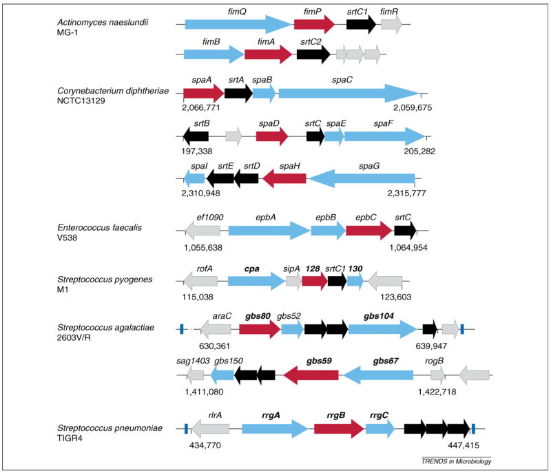
Pilus gene operons. Graphic presentation of pilus gene clusters identified in the chromosome of Actinomyces naeslundii MG-1, Corynebacterium diphtheriae NCTC13129, Enterococcus faecalis V538, Streptococcus pyogenes M1, Streptococcus agalactiae 2603V/R and Streptococcus pneumoniae TIGR4. Each cluster contains pilus-specific sortase gene(s) (black), genes encoding a major subunit (red) and minor pilins (aqua). Some of the clusters are flanked by transposable elements (blue). Genes encoding pilins used in vaccine studies are shown in bold. Uncharacterized genes are colored in gray. Numbers below clusters indicate the genomic location of pilus gene clusters.

Model of pilus biogenesis. Pilin precursors (SpaA, denoted by pink circles; SpaB, denoted by dark-aqua ovals; and SpaC, denoted by light-aqua ovals) are synthesized in the cytoplasm and translocated across the membrane by the Sec machinery (step 1). At the exoplasm, the precursors subsequently form acyl–enzyme intermediates with the housekeeping sortase (green) (step 2) or pilus-specific sortase (gray). These enzyme intermediates are capable of transferring these pilins to the lipid II precursor, thus anchoring monomeric pilins to the cell wall (step 3 in A). The pilus-specific sortase catalyzes pilus polymerization (step 4) by the mechanism described in Box 2. Pilus polymerization is terminated when pilus polymers are transferred to lipid II in one of two possible ways. In one pathway, the housekeeping sortase having a SpaA monomer would receive the pilus polymer from the pilus-specific sortase (step 5) and transfer the polymer to lipid II (step 6). In the alternative pathway (not shown), the pilus-specific sortase would transfer the polymer directly to lipid II. Red diamonds denote the D-diaminopimelic moiety of the cell wall pentapeptide. SecYEG stands for the three subunits of the general secretion machinery (Sec).
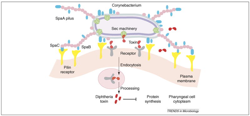
Model of pilus-mediated adhesion and pathogenesis. This general model applies to various Gram-positive pathogens. We depict Corynebacterium diphtheriae as a specific case. Adhesive fibers make initial contact with host cell receptors, whereas cell-wall-linked pilins mediate the formation of an intimate zone of adhesion. This enables additional ligand–receptor interactions, the efficient delivery of virulence factors, and the intracellular invasion of certain pathogens.
References
-
- Sjöquist J, et al. Protein A isolated from Staphylococcus aureus after digestion with lysostaphin. Eur J Biochem. 1972;29:572–578. - PubMed
-
- Sjöquist J, et al. Localization of protein A in the bacteria. Eur J Biochem. 1972;30:190–194. - PubMed
-
- Schneewind O, et al. Sorting of protein A to the staphylococcal cell wall. Cell. 1992;70:267–281. - PubMed
-
- Mazmanian SK, et al. Staphylococcus aureus sortase, an enzyme that anchors surface proteins to the cell wall. Science. 1999;285:760–763. - PubMed
Publication types
MeSH terms
Substances
Grants and funding
LinkOut - more resources
Full Text Sources
Other Literature Sources
Molecular Biology Databases
