Review
. 2007 Dec 17;7(1):224-33.
doi: 10.1102/1470-7330.2007.0032.

MDCT of small bowel tumours

Affiliations
Review

MDCT of small bowel tumours

Johannes Sailer et al. Cancer Imaging. .

Abstract

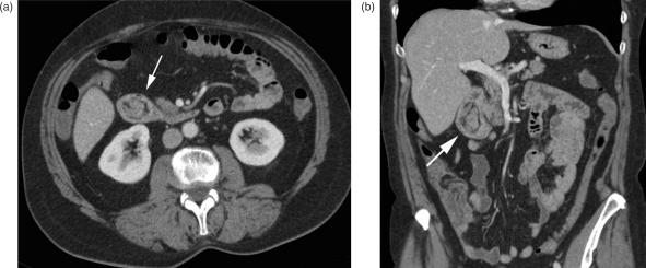
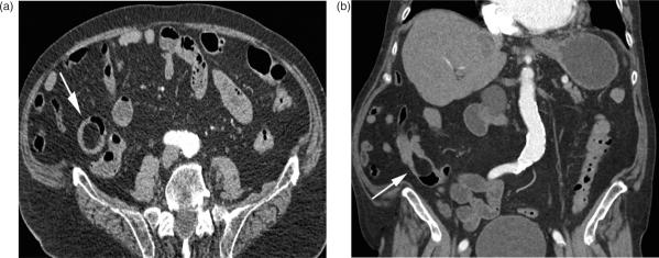
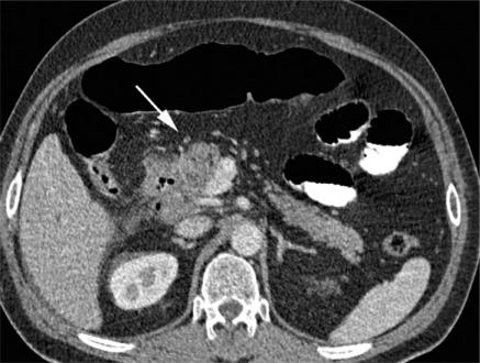
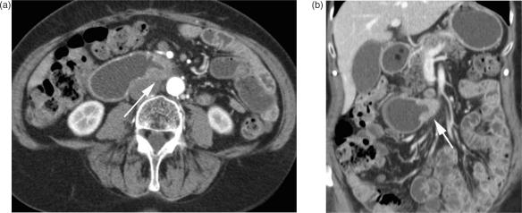
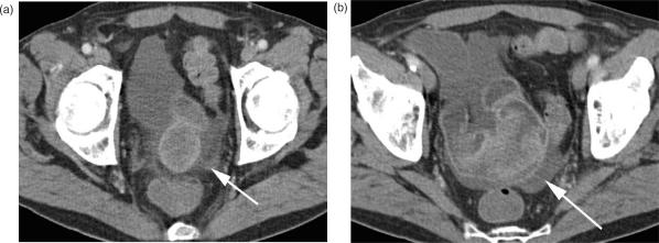
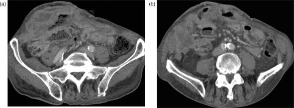
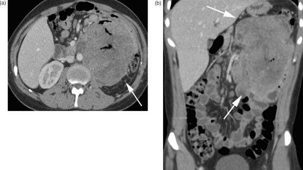
Primary benign and malignant neoplasm of the small bowel are rare. Malignant tumours often present late symptoms resulting in a poor prognosis. Early detection of small bowel neoplasms is desirable but challenging for both clinicians and radiologists. Conventional double contrast enteroclysis was the method of choice in small bowel imaging but is increasingly being replaced by cross-sectional imaging methods as computed tomography (CT) and magnetic resonance imaging (MRI). Multidetector CT (MDCT) produces high-resolution cross-sectional imaging of the abdomen and the small bowel. It allows multiplanar visualisation of small bowel tumours, demonstrates signs of small bowel obstruction as well as the mural and extramural extent of small bowel malignancies. This aids planning for surgical resection. In addition, liver metastases or peritoneal seeding can be detected with CT. The best visualisation of small bowel neoplasms is achieved with CT enteroclysis or enterography and this review discusses these techniques and MDCT characteristics of small bowel tumours.


Figures

Figure 1
Figure 1
Duodenal adenoma. (a) Axial MDCT of a polypoid intraluminal mass (arrow) in the duodenum with distinct contrast enhancement. A fat rim allows good delineation of the bowel wall, there are no signs of bowel wall infiltration. (b) Coronal and (c) sagittal reformation.
Figure 2
Figure 2
Lipoma of the small intestine. (a) Axial and (b) coronal MDCT images of an intraluminal mass (arrow) in the terminal ileum with fat-equivalent attenuation values.
Figure 3
Figure 3
Duodenal adenocarcinoma. MDCT shows a contrast-enhanced mass of the descending part of the duodenum infiltrating the surrounding mesenterial fat and the pancreas head (arrow). It is difficult to differentiate between duodenal cancer invading the pancreatic head and (the much more common situation of) pancreatic cancer invading the duodenum.
Figure 4
Figure 4
Duodenal adenocarcinoma (horizontal part). (a) Axial and (b) coronal CT images with contrast-enhanced circumferential wall thickening of the horizontal part of the duodenum (arrow) causing stenosis with prestenotic dilatation, typical for adenocarcinoma.
Figure 5
Figure 5
Neuroendocrine tumour (NET). (a) Axial MDCT image with contrast-enhanced focal nodular wall thickening of the ileum (arrow); (b) the desmoplastic reaction within the adjacent mesenterial fat with typical calcifications below (arrow); (c) coronal reformation.
Figure 6
Figure 6
Small bowel lymphoma. (a) Segmental wall thickening of an ileal loop (arrow). Typically there are no signs of luminal narrowing. (b) The same lesion on coronal reformation.
Figure 7
Figure 7
Gastrointestinal stroma tumour (GIST). (a) A contrast-enhanced ileal mass in the lower abdomen, infiltrating into the mesenteric fat (arrow). Adjacent to the mass, there are small extraluminal air bubbles as a sign of perforation; mesenteric stranding indicates local peritonitis. (b) Coronal reformation and small mesenteric lymph nodes.
Figure 8
Figure 8
Low grade GIST of the jejunum. (a) Axial and (b) coronal MDCT images of another patient with a large rim-enhanced mass in the jejunum histologically verified as low grade GIST.
Figure 9
Figure 9
Leiomyosarcoma of the small bowel. (a,b) Axial MDCT images of the lower abdomen with segmental contrast-enhanced wall thickening, histologically verified as a leiomyosarcoma. The tumour is mostly located intraluminal; distinct perifocal stranding suggests mesenteric infiltration.
Figure 10
Figure 10
Peritoneal carcinomatosis. (a,b) Extensive peritoneal seeding in a patient with end-stage ovarian carcinoma. There are multifocal contrast enhancing nodular masses infiltrating the small bowel wall and the ventral abdominal wall.
Figure 11
Figure 11
Small bowel metastasis. (a) Axial and (b) coronal CT images of a large mass in the left lower part of the abdomen. Small central air bubbles are the only sign of an intestinal origin. The mass was verified as a metastasis of a seminoma.

Similar articles

Cited by

References

    1. Good CA. Tumors of the small intestine. AJR. 1963;89:685–705. - PubMed
    1. Ashley SW, Wells SA. Tumors of the small intestine. Semin Oncol. 1988;15:116–28. - PubMed
    1. Wilson JM, Melvin DB, Gray GF, et al. Primary malignancies of the small bowel: a report of 96 cases and review of the literature. Ann Surg. 1974;180:175–79. - PMC - PubMed
    1. Lewis BS, Kornbluth A. Small bowel tumors: yield of enteroscopy. Gut. 1991;32:763–5. - PMC - PubMed
    1. May A, Nachbar L, Schneider M, Ell C. Double-balloon enteroscopy (push-and-pull enteroscopy) of the small bowel: feasibility, diagnostic and therapeutic yield in patients with suspected small bowel disease. Gastrointest Endosc. 2005;62:62–70. - PubMed