. 2008 Feb;146(2):589-601.
doi: 10.1104/pp.107.108183. Epub 2007 Dec 14.

The Arabidopsis AtOPT3 protein functions in metal homeostasis and movement of iron to developing seeds

Affiliations

The Arabidopsis AtOPT3 protein functions in metal homeostasis and movement of iron to developing seeds

Minviluz G Stacey et al. Plant Physiol. 2008 Feb.

Abstract

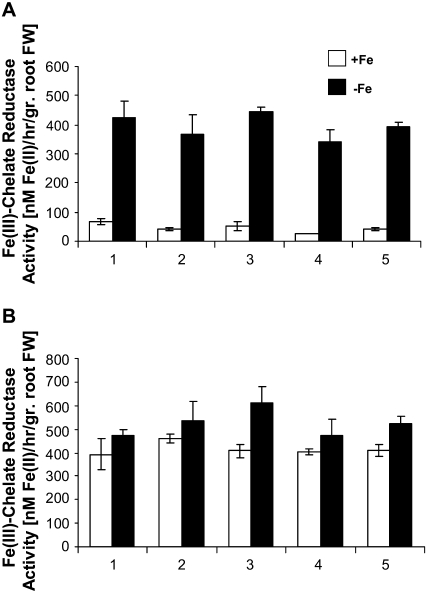
The Arabidopsis thaliana AtOPT3 belongs to the oligopeptide transporter (OPT) family, a relatively poorly characterized family of peptide/modified peptide transporters found in archebacteria, bacteria, fungi, and plants. A null mutation in AtOPT3 resulted in embryo lethality, indicating an essential role for AtOPT3 in embryo development. In this article, we report on the isolation and phenotypic characterization of a second AtOPT3 mutant line, opt3-2, harboring a T-DNA insertion in the 5' untranslated region of AtOPT3. The T-DNA insertion in the AtOPT3 promoter resulted in reduced but sufficient AtOPT3 expression to allow embryo formation in opt3-2 homozygous seeds. Phenotypic analyses of opt3-2 plants revealed three interesting loss-of-function phenotypes associated with iron metabolism. First, reduced AtOPT3 expression in opt3-2 plants resulted in the constitutive expression of root iron deficiency responses regardless of exogenous iron supply. Second, deregulation of root iron uptake processes in opt3-2 roots resulted in the accumulation of very high levels of iron in opt3-2 tissues. Hyperaccumulation of iron in opt3-2 resulted in the formation of brown necrotic areas in opt3-2 leaves and was more pronounced during the seed-filling stage. Third, reduced AtOPT3 expression resulted in decreased accumulation of iron in opt3-2 seeds. The reduced accumulation of iron in opt3-2 seeds is especially noteworthy considering the excessively high levels of accumulated iron in other opt3-2 tissues. AtOPT3, therefore, plays a critical role in two important aspects of iron metabolism, namely, maintenance of whole-plant iron homeostasis and iron nutrition of developing seeds.


Figures

Figure 1.
Figure 1.
Isolation of a T-DNA insertion mutation in the promoter of AtOPT3. A, Diagram of the T-DNA and insertion site upstream of the AtOPT3 gene in opt3-2. The T-DNA is inserted 36 bp upstream of the AtOPT3 start codon in the orientation indicated. Boxes represent exons and lines represent introns. RB, Right border; LB, left border. Bar = 500 bp. B, Semiquantitative determination of AtOPT3 expression in Col-0 and opt3-2 by RT-PCR. Plants were grown on Murashige and Skoog plates for 15 d then grown for 4 d under Fe-sufficient (+) or Fe-deficient (−) conditions. Total RNA was isolated from roots and shoots and RT-PCR amplification of AtOPT3 is shown. As control, RT-PCR amplification of Actin2 is also shown.
Figure 2.
Figure 2.
Phenotypic characterization of opt3-2 seedlings. A, Col-0 and opt3-2 seedlings grown under Fe-sufficient (+Fe) or Fe-deficient (−Fe) conditions. opt3-2 seedlings were slightly smaller than the wild type but showed no obvious chlorosis when grown under Fe-sufficient condition. Both opt3-2 and the wild type turned chlorotic when grown under Fe deficiency for 4 d. Bar = 2 mm. B, Localization of Fe3+ in leaves (top) and roots (bottom) of Col-0 and opt3-2 seedlings grown under Fe-sufficient condition. Fe3+ localization was determined using Perl's stain. Arrows identify Fe3+ staining. Bar = 150 μm. C, Transverse sections of Col-0 and opt3-2 leaves. Subepidermal layer of columnar palisade parenchyma cells was observed in Col-0 leaves but was lacking in opt3-2 leaves. Palisade parenchyma cells in opt3-2 leaves failed to differentiate and remained indistinguishable from the underlying spongy parenchyma cells. EP, Epidermis; PP, palisade parenchyma; SP, spongy parenchyma; UPP, undifferentiated palisade parenchyma. Bar = 50 μm.
Figure 3.
Figure 3.
Constitutive expression of Fe deficiency responses in opt3-2 roots. A, Fe(III)-chelate reductase activity of Col-0 and opt3-2 plants under Fe-sufficient (+Fe) and Fe-deficient (−Fe) conditions. Means and se of 12 replicates done over two trials are shown. Value for each replicate represents activity obtained from three pooled plants. B, Determination of AtIRT1 and AtFER1 expression in Col-0 and opt3-2 by semiquantitative RT-PCR. Total RNA was isolated from roots and shoots of Col-0 and opt3-2 plants grown for 4 d under Fe-sufficient (+) or Fe-deficient (−) conditions. RT-PCR amplification of AtIRT1 and AtFER1 transcripts is shown. As control, RT-PCR amplification of Actin2 is also shown. Numbers below each lane indicate the relative intensity of the AtFER1 band to the corresponding Actin2 band. Comparable patterns of AtFER1 expression were obtained in a second biological trial (data not shown).
Figure 4.
Figure 4.
Genetic complementation of the constitutively expressed root Fe(III)-chelate reductase activity in opt3-2 plants. Fe3+-chelate reductase activity assays were performed on five independent transgenic lines transformed with AtOPT3 (A) or GUS (B) expressed from the AtOPT3 promoter. Plants were grown under Fe-sufficient (+FE) or Fe-deficient (−Fe) conditions for 4 d. For each line, means and se of three replicates are shown.
Figure 5.
Figure 5.
Metal and macronutrient concentrations in Col-0 and opt3-2 plants at bolting. Plants were grown in soil until bolting, then aerial tissues from 16 plants were harvested, pooled, and analyzed for metal (A) and macronutrient (B) concentration. Means and se obtained from three biological replicates are shown.
Figure 6.
Figure 6.
Overaccumulation of Fe, Mn, Zn, and Cu in opt3-2 plants at 10 d after bolting. Rosette leaves (A), inflorescence stems (B), and siliques (C) were harvested at 10 d after bolting and analyzed for metal concentration. Samples represent pooled tissues from 16 plants. At 10 d after bolting, very few flower buds remained and siliques were not yet dehiscent. Inflorescence stems included cauline leaves and a few remaining flowers and excluded siliques that were detached and assayed separately. Means and se obtained from three biological replicates are shown.
Figure 7.
Figure 7.
Leaf necrosis and high levels of stainable Fe3+ in various tissues of opt3-2 plants. A to C, Mature leaves from opt3-2 (A, B) and Col-0 (C) plants at 3 d after bolting. Arrows (A, B) indicate necrotic lesions in opt3-2 leaves. D to P, Histochemical localization of Fe3+ in leaves (D–F), flowers (G–J), and siliques (K–P) of Col-0 (D, G, K–M) and opt3-2 (E, F, H–J, N–P) plants. Arrows indicate strong Fe3+ staining in anther (J), distal ends of siliques (N, O), and vascular tissues of seed coat and funiculus (P) of opt3-2 plants. Q and R, Representative Col-0 (Q) and opt3-2 (R) siliques at desiccation stage (stage 19). Compared to Col-0, opt3-2 siliques appeared grayish-brown and shriveled at distal ends. Bar = 1 mm in A, C, D, and E; 300 μm in B, M, and P; 150 μm in F; 500 μm in G to J; 2 mm in K, L, N, P, Q, and R.
Figure 8.
Figure 8.
Reduced stored Fe in opt3-2 seeds. A, Metal concentration in Col-0 and opt3-2 seeds. Compared with the wild type, opt3-2 seeds showed lower concentration of Fe and higher concentration of Mn, Zn, and Cu. B, Macronutrient concentration in Col-0 and opt3-2 seeds. No significant difference in Ca, K, Mg, and P concentration in wild-type and opt3-2 seeds was observed. Means and se obtained from three biological replicates are shown.
Figure 9.
Figure 9.
Reduced stainable Fe3+ in developing opt3-2 seeds and reduced growth of opt3-2 seedlings under Fe-starved conditions. A, Localization of Fe3+ in developing embryos of Col-0 and opt3-2 seeds at early (excised embryos, top) and late (bottom) curled-cotyledon stages of embryo development. Arrows indicate stored Fe in axis and cotyledons of Col-0 embryo. Perl's staining to visualize Fe3+ was done for 30 min. Arrows indicate Fe3+ in embryonic vasculature. Bars = 120 μm. B, Localization of Fe3+ in excised opt3-2 embryos after 30 (left and middle) and 60 (right) min of Perl's staining. Similar to Col-0, longer staining period showed that Fe3+ was localized to developing vasculature of opt3-2 embryos. Arrows indicate Fe3+ in embryonic vasculature. Bars = 120 μm. C, Seedling growth of Col-0 and opt3-2 under Fe-starved condition. Seeds were germinated and grown horizontally for 8 d on medium lacking Fe. Bars = 5 mm.
Figure 10.
Figure 10.
Reduced seed yield in opt3-2. A, Seed yield per plant. Means and se obtained for 10 Col-0 or 10 opt3-2 plants are shown. B, Representative fully extended siliques from Col-0 and opt3-2 plants. Siliques were obtained from approximately similar positions on the main inflorescence stems of Col-0 and opt3-2 at 10 d after bolting. Bar = 4 mm. C, Representative green and desiccated Col-0 and opt3-2 seeds. Bars = 500 μm (top) and 300 μm (bottom).

Similar articles

Cited by

References

    1. Alonso JM, Stepanova AN, Leisse TJ, Kim CJ, Chen H, Shinn P, Stevenson DK, Zimmerman J, Barajas P, Cheuk R, et al (2003) Genome-wide insertional mutagenesis of Arabidopsis thaliana. Science 301 653–657 - PubMed
    1. Bashir K, Inoue H, Nagasaka S, Takahashi M, Nakanishi H, Mori S, Nishizawa NK (2006) Cloning and characterization of deoxymugineic acid synthase genes from graminaceous plants. J Biol Chem 281 32395–32402 - PubMed
    1. Bechtold N, Pelletier G (1998) In planta Agrobacterium-mediated transformation of adult Arabidopsis thaliana plants by vacuum infiltration. Methods Mol Biol 82 259–266 - PubMed
    1. Bellaoui M, Keddie JS, Gruissem W (2003) DCL is a plant-specific protein required for plastid ribosomal RNA processing and embryo development. Plant Mol Biol 53 531–543 - PubMed
    1. Bourbouloux A, Shahi P, Chakladar A, Delrot S, Bachhawat AK (2000) Hgt1p, a high affinity glutathione transporter from the yeast Saccharomyces cerevisiae. J Biol Chem 275 13259–13265 - PubMed

Publication types

MeSH terms