. 2008 Feb;190(4):1335-43.
doi: 10.1128/JB.01764-07. Epub 2007 Dec 14.

Differential regulation of high-affinity phosphate transport systems of Mycobacterium smegmatis: identification of PhnF, a repressor of the phnDCE operon

Affiliations

Differential regulation of high-affinity phosphate transport systems of Mycobacterium smegmatis: identification of PhnF, a repressor of the phnDCE operon

Susanne Gebhard et al. J Bacteriol. 2008 Feb.

Abstract

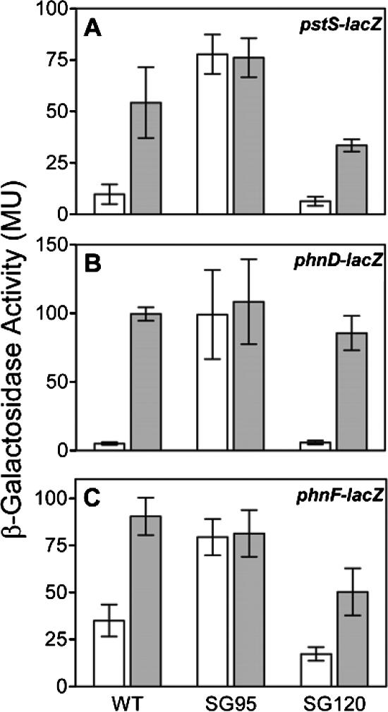
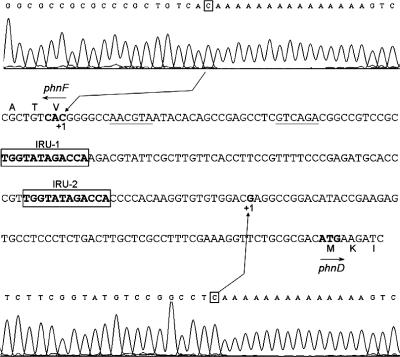
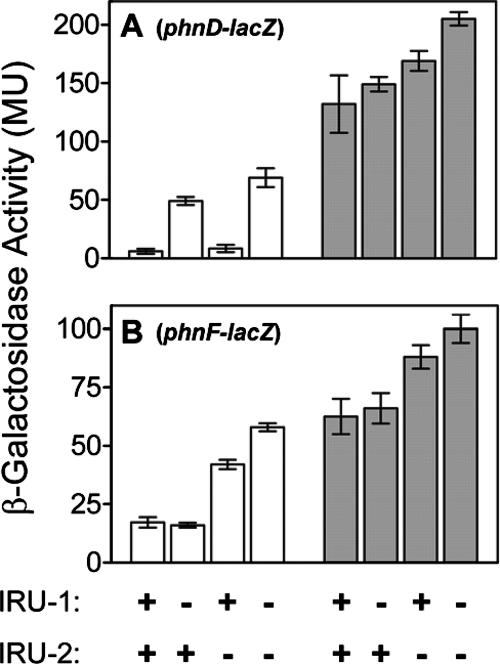
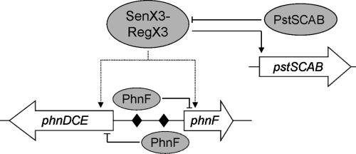
The uptake of phosphate into the cell via high-affinity, phosphate-specific transport systems has been studied with several species of mycobacteria. All of these species have been shown to contain several copies of such transport systems, which are synthesized in response to phosphate limitation. However, the mechanisms leading to the expression of the genes encoding these transporters have not been studied. This study reports on the investigation of the regulation of the pstSCAB and the phnDCE operons of Mycobacterium smegmatis. The phn locus contains an additional gene, phnF, encoding a GntR-like transcriptional regulator. Expression analyses of a phnF deletion mutant demonstrated that PhnF acts as a repressor of the phnDCE operon but does not affect the expression of pstSCAB. The deletion of pstS, which is thought to cause the constitutive expression of genes regulated by the two-component system SenX3-RegX3, led to the constitutive expression of the transcriptional fusions pstS-lacZ, phnD-lacZ, and phnF-lacZ, suggesting that phnDCE and phnF are conceivably new members of the SenX3-RegX3 regulon of M. smegmatis. Two presumptive binding sites for PhnF in the intergenic region between phnD and phnF were identified and shown to be required for the repression of phnD and phnF, respectively. We propose a model in which the transcription of pstSCAB is controlled by the two-component SenX3-RegX3 system, while phnDCE and phnF are subject to dual control by SenX3-RegX3 and PhnF.


Figures

FIG. 1.
FIG. 1.
Sequence analysis of M. smegmatis PhnF. (A) Map of the phn genes of M. smegmatis. The names of the loci, as annotated in the provisional genome sequence of M. smegmatis mc2155, are indicated below the arrows. (B) Alignment of PhnF proteins from E. coli (Eco) and M. smegmatis (Msm). Identities are shown in black, and similarities (threshold, 90%) are shown in a gray background. The predicted secondary structure of M. smegmatis PhnF is shown below the sequence, with H indicating an α helix and E indicating a β sheet. Triangles indicate the large (L) and polar (H or T) signature residues of HutC subfamily proteins (2). Diamonds indicate the residues conserved among HutC members, which delineate the binding site of E. coli PhnF (8). Solid and open symbols show residues conserved and not conserved, respectively, in M. smegmatis PhnF.
FIG. 2.
FIG. 2.
Transcriptional activities of phnF-lacZ during phosphate-limited growth. Cells were grown in modified minimal Sauton's medium (5 g glycerol liter−1, 4 g l-asparagine liter−1, 200 μM Pi) and monitored for growth, expressed as OD600 (▪), phosphate concentration in the medium (Pi) (▵), and β-galactosidase activity (β-Gal), expressed as MU (□). Representative results of two independent experiments are shown.
FIG. 3.
FIG. 3.
Allelic replacement of phnF. (A) Schematic diagram (not drawn to scale) of allelic replacement of phnF with aphA-3. SmaI restriction sites and band sizes as detected in panel B are indicated. The bold line shows the fragment used as a probe. (B) Southern hybridization analysis of the replacement of phnF in strain SG62. SmaI-digested genomic DNA from the wild-type (WT) strain and from strain SG62 (ΔphnF) was probed with the radiolabeled left-flank PCR product of the deletion construct. Molecular sizes are indicated in kilobases.
FIG. 4.
FIG. 4.
Dot blot analysis of RNA from wild-type and mutant strains. Fourfold dilutions of total RNA isolated from cells grown in high-phosphate medium (100 mM; +) or subjected to phosphate starvation for 2 h (−) were spotted onto nylon membranes. Membranes were probed with radiolabeled PCR products from internal fragments of pstC (A, top panel) or phnC (A, bottom panel, and B). Amounts of total RNA per spot are shown in ng. Strains are indicated above the autoradiographs. (A) WT, wild-type; ΔphnF, phnF deletion strain SG62; cphnF, phnF-complemented strain SG111. (B) WT, wild-type; ΔpstS, pstS deletion strain SG95; cpstS, pstS-complemented strain SG120. Representative results of two to three independent experiments are shown. Below each panel, 16S and 23S rRNA bands from 300 ng total RNA per sample on an agarose gel stained with ethidium bromide are shown as controls. Lanes correspond to the samples in the rows above.
FIG. 5.
FIG. 5.
Expression levels of pstS-lacZ, phnD-lacZ, and phnF-lacZ in various genetic backgrounds of M. smegmatis. Cells of the wild type (WT), the pstS deletion strain (SG95), and the complemented strain (SG120) carrying various lacZ fusion constructs were grown in ST medium containing 100 mM Pi (open bars) or subjected to phosphate starvation for 2 h (gray bars). β-Galactosidase activities are given as MU. (A) Cells harboring the pSG42 plasmid (pstS-lacZ). (B) Cells harboring the pSG10 plasmid (phnD-lacZ). (C) Cells harboring the pSG18 plasmid (phnF-lacZ). Results are shown as the means and standard deviations of results from two to four independent experiments. Differences between cells grown in 100 mM Pi and phosphate-starved cells of the wild-type and those of the SG120 strain and differences between the wild-type cells grown in 100 mM Pi and the cells of the SG95 strain are statistically significant (P < 0.05).
FIG. 6.
FIG. 6.
Sequence analysis of the intergenic region between phnF and phnD. Transcriptional start sites for phnF and phnD were determined by 5′ RACE analysis as shown in the top and bottom panels, respectively (traces are given as reverse sequences). The sequence given in the middle panel shows a region of the coding strand for phnD, encompassing the first three codons of phnF and phnD (the first three amino acids are indicated) and the intergenic region. It should be noted that the sequence for phnF is therefore given as a reverse sequence. Start codons are shown in bold; transcriptional start sites are shown in bold and are indicated as +1. Putative −10 and −35 regions for phnF are underlined. The two IRUs, which constitute presumptive PhnF binding sites, are boxed.
FIG. 7.
FIG. 7.
Effects of site-directed mutagenesis of the IRUs in the intergenic region between phnF and phnD on gene expression. Changes were introduced into IRU-1 and IRU-2 (see text for details), and effects were monitored as the expression of phnD-lacZ (A) and phnF-lacZ (B) transcriptional fusions. Cells were grown in minimal Sauton's medium containing 100 mM phosphate (open bars) or starved in phosphate-free medium for 2 h (gray bars). The presence of wild-type sequences (+) or site-directed changes (−) in each IRU are shown below the graphs. Results are shown as the means and standard deviations from three independent experiments.
FIG. 8.
FIG. 8.
Model for the regulation of pstSCAB, phnDCE, and phnF in M. smegmatis. Genes are shown as open arrows; proteins are shown as gray ovals. Flat-headed arrows indicate negative regulation; a pointed arrowhead indicates positive regulation. Dotted lines indicate that the proposed regulation may be direct or indirect. The presumptive PhnF binding sites are shown as black diamonds.

Similar articles

Cited by

References

    1. Agarwal, N., and A. K. Tyagi. 2006. Mycobacterial transcriptional signals: requirements for recognition by RNA polymerase and optimal transcriptional activity. Nucleic Acids Res. 344245-4257. - PMC - PubMed
    1. Aravind, L., and V. Anantharaman. 2003. HutC/FarR-like bacterial transcription factors of the GntR family contain a small molecule-binding domain of the chorismate lyase fold. FEMS Microbiol. Lett. 22217-23. - PubMed
    1. Bardin, S., S. Dan, M. Osteras, and T. M. Finan. 1996. A phosphate transport system is required for symbiotic nitrogen fixation by Rhizobium meliloti. J. Bacteriol. 1784540-4547. - PMC - PubMed
    1. Cuff, J. A., M. E. Clamp, A. S. Siddiqui, M. Finlay, and G. J. Barton. 1998. JPred: a consensus secondary structure prediction server. Bioinformatics 14892-893. - PubMed
    1. Gebhard, S., S. L. Tran, and G. M. Cook. 2006. The Phn system of Mycobacterium smegmatis: a second high-affinity ABC-transporter for phosphate. Microbiology 1523453-3465. - PubMed

Publication types

MeSH terms

LinkOut - more resources