. 2008 Feb;190(4):1224-36.
doi: 10.1128/JB.01780-07. Epub 2007 Dec 14.

Genetic and biochemical analysis of CodY-binding sites in Bacillus subtilis

Affiliations

Genetic and biochemical analysis of CodY-binding sites in Bacillus subtilis

Boris R Belitsky et al. J Bacteriol. 2008 Feb.

Abstract

CodY is a global transcriptional regulator that is known to control directly the expression of at least two dozen operons in Bacillus subtilis, but the rules that govern the binding of CodY to its target DNA have been unclear. Using DNase I footprinting experiments, we identified CodY-binding sites upstream of the B. subtilis ylmA and yurP genes. The protected regions overlapped versions of a previously proposed CodY-binding consensus motif, AATTTTCWGAAAATT. Multiple single mutations were introduced into the CodY-binding sites of the ylmA, yurP, dppA, and ilvB genes. The mutations affected both the affinity of CodY for its binding sites in vitro and the expression in vivo of lacZ fusions that carry these mutations in their promoter regions. Our results show that versions of the AATTTTCWGAAAATT motif, first identified for Lactococcus lactis CodY, with up to five mismatches play an important role in the interaction of B. subtilis CodY with DNA.


Figures

FIG. 1.
FIG. 1.
(A) Primer extension analysis of the ylmA mRNA. Primer oBB102 annealing to the lacZ gene of the ylmA-lacZ fusion was extended with reverse transcriptase by using as the template total RNAs from fusion-containing strains BB2718 (codY+, lane 1) and BB2724 (codY, lane 2) grown in glucose-ammonium medium containing 16 aa. The sequence of the template strand of pBB1485 determined from reactions primed with oBB102 is shown to the right. The apparent transcription start site of the ylmA gene is in bold and marked by the +1 notation. The bent arrow indicates the direction of transcription. (B) Primer extension analysis of the yurP mRNA. Primer oBB102 annealing to the lacZ gene of the yurP-lacZ fusion was extended with reverse transcriptase by using as the template total RNA from fusion-containing strains BB2675 (codY+, lane 1) and BB2687 (codY, lane 2) grown in glucose-ammonium medium containing 16 aa. The sequence of the template strand of pBB1468 determined from reactions primed with oBB102 is shown to the left. The apparent transcription start site of the yurP gene is in bold and marked by the +1 notation. The bent arrow indicates the direction of transcription.
FIG. 2.
FIG. 2.
DNase I footprinting analysis of CodY binding to the ylmA regulatory region. (A) The ylmAp+ DNA fragment labeled on the template strand was incubated with increasing amounts of purified CodY in the presence of ILV and GTP and then with DNase I. The sequence of the ylmA region was determined with pBB1485 as the template and oBB253 as the primer and is shown to the right. The apparent transcription start site and direction of ylmA transcription are shown by the bent arrow. The junction between ylmA and vector DNA is indicated by the triangle. The protected area is indicated by the vertical line. The CodY concentration used (nM monomer) is indicated above each lane. (B) The ylmAp+, ylmAp1, or ylmAp2 DNA fragment labeled on the template strand was incubated with purified CodY in the presence of ILV and GTP and then with DNase I. The protected area is indicated by the vertical line. The CodY concentration used (nM monomer) is indicated above each lane. (C) The ylmAp1 DNA fragment labeled on the template strand was incubated with purified CodY in the absence of ILV and GTP and then with DNase I. The region protected by CodY when ILV and GTP are present is indicated by the vertical line. The CodY concentration used (nM monomer) is indicated above each lane. (D) Sequence of the coding (nontemplate) strand of the ylmA regulatory region. The −10 and −35 promoter regions, transcription start site, and CodY-binding motif are in bold. The direction of translation is indicated by the arrow. The likely initiation codon and sequence protected by CodY in DNase I footprinting experiments on the template strand of DNA are underlined. The downstream terminus of the ylmA DNA fragment used to create the lacZ fusion is indicated by the triangle.
FIG. 3.
FIG. 3.
Stimulation by GTP and ILV of CodY binding to the ylmA promoter. The ylmAp+ DNA fragment labeled on the template strand was incubated with purified CodY in the absence of the effectors or in the presence of GTP, ILV, or both and then with DNase I. The protected area is indicated by the vertical line. The CodY concentration used (nM monomer) is indicated above each lane.
FIG. 4.
FIG. 4.
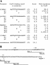
CodY-binding motifs and mutations. (A) Mismatches with the consensus sequence are indicated by lowercase letters; the number of mismatches is in parentheses. Mutations are shown below the sequence lanes as substituting nucleotides. For dppA, the sequence shown corresponds to dppA box 3 in Table 3. The scores for individual CodY-binding motifs were generated with the help of the software package at http://rsat.ulb.ac.be/rsat/ by using the position-specific weight matrix and reflect the similarity of these motifs to the entire set of 27 CodY-binding motifs used to create the matrix. Fold regulation by CodY reflects differences in the expression of the corresponding lacZ fusions between the wild-type and codY null strains in 16-aa-containing medium. (B) Locations of putative CodY-binding motifs in the dppA regulatory region. The −10 and −35 promoter regions and transcription start site are in bold. dppA box I, box II, box III, and box IV are underlined. The uppercase letters correspond to the region protected by CodY in DNase I footprinting experiments with the wild-type dppA promoter (26, 52). The vertical arrow indicates the site of a 4 bp-insertion within the dppAp49 allele. The 12-bp dppAp129 deletion affects nucleotides in the positions from +5 to +16.
FIG. 5.
FIG. 5.
DNase I footprinting analysis of CodY binding to the yurP regulatory region. (A) The yurPp+ DNA fragment labeled on the template strand was incubated with purified CodY in the presence of ILV and GTP and then with DNase I. The sequence of the yurP region was determined with pBB1468 as the template and oBB102 as the primer and is shown to the right. The apparent transcription start site and direction of yurP transcription are shown by the bent arrow. The protected area is indicated by the vertical line. The CodY concentration used (nM monomer) is indicated above each lane. (B) The yurPp+, yurPp1, or yurPp3 DNA fragment labeled on the template strand was incubated with purified CodY in the presence of ILV and GTP and then with DNase I. The protected area is indicated by the vertical line. The CodY concentration used (nM monomer) is indicated above each lane. (C) The yurPp+ DNA fragment labeled on the template strand was incubated with purified CodY in the absence of ILV and GTP and then with DNase I. The region protected by CodY when ILV and GTP are present is indicated by the vertical line. The CodY concentration used (nM monomer) is indicated above each lane. (D) Sequence of the yurP regulatory region. The −10 and −35 promoter regions, transcription start site, and CodY-binding motif are in bold. The direction of translation is indicated by the arrow. The likely initiation codon and sequence protected by CodY in DNase I footprinting experiments on the template strand of DNA are underlined.
FIG. 6.
FIG. 6.
Position-specific weight matrix of CodY-binding motifs. Shown is the position-specific weight matrix for 27 established and likely B. subtilis CodY-binding motifs showing the relative frequency of occurrence of each of the four nucleotides (A, T, C, or G) at each of the 15 positions of the motif. The logo presentation of the matrix (50) was created at http://weblogo.berkeley.edu/.

Similar articles

Cited by

References

    1. Altschul, S. F., T. L. Madden, A. A. Schaffer, J. Zhang, Z. Zhang, W. Miller, and D. J. Lipman. 1997. Gapped BLAST and PSI-BLAST: a new generation of protein database search programs. Nucleic Acids Res. 253389-3402. - PMC - PubMed
    1. Atkinson, M. R., L. V. Wray, Jr., and S. H. Fisher. 1990. Regulation of histidine and proline degradation enzymes by amino acid availability in Bacillus subtilis. J. Bacteriol. 1724758-4765. - PMC - PubMed
    1. Belitsky, B. R., M. C. Gustafsson, A. L. Sonenshein, and C. Von Wachenfeldt. 1997. An lrp-like gene of Bacillus subtilis involved in branched-chain amino acid transport. J. Bacteriol. 1795448-5457. - PMC - PubMed
    1. Belitsky, B. R., and A. L. Sonenshein. 2002. GabR, a member of a novel protein family, regulates utilization of γ-aminobutyrate in Bacillus subtilis. Mol. Microbiol. 45569-583. - PubMed
    1. Belitsky, B. R., and A. L. Sonenshein. 1998. Role and regulation of Bacillus subtilis glutamate dehydrogenase genes. J. Bacteriol. 1806298-6305. - PMC - PubMed

Publication types

MeSH terms

LinkOut - more resources