Comparative Study
. 2008 Apr;190(8):2804-13.
doi: 10.1128/JB.01572-07. Epub 2007 Dec 14.

In vivo growth of Pseudomonas aeruginosa strains PAO1 and PA14 and the hypervirulent strain LESB58 in a rat model of chronic lung infection

Affiliations
Comparative Study

In vivo growth of Pseudomonas aeruginosa strains PAO1 and PA14 and the hypervirulent strain LESB58 in a rat model of chronic lung infection

Irena Kukavica-Ibrulj et al. J Bacteriol. 2008 Apr.

Abstract

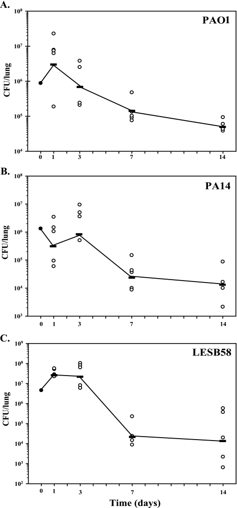
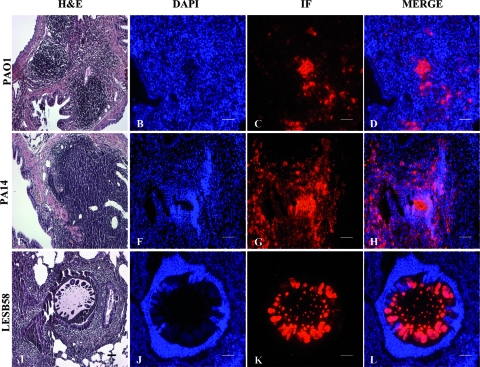
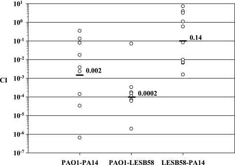
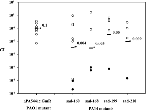
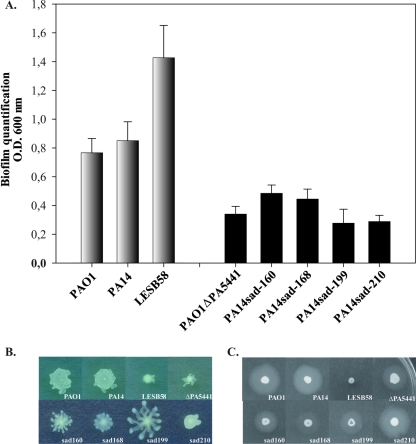
Pseudomonas aeruginosa chronic lung infections are the major cause of morbidity and mortality in cystic fibrosis (CF) patients. The P. aeruginosa strains PAO1 and PA14 were compared with the Liverpool epidemic strain LESB58 to assess in vivo growth, infection kinetics, and bacterial persistence and localization within tissues in a rat model of chronic lung infection. The three P. aeruginosa strains demonstrated similar growth curves in vivo but differences in tissue distribution. The LESB58 strain persisted in the bronchial lumen, while the PAO1 and PA14 strains were found localized in the alveolar regions and grew as macrocolonies after day 7 postinfection. Bacterial strains were compared for swimming and twitching motility and for the production of biofilm. The P. aeruginosa LESB58 strain produced more biofilm than PAO1 and PA14. Competitive index (CI) analysis of PAO1, PA14, and LESB58 in vivo indicated CI values of 0.002, 0.0002, and 0.14 between PAO1-PA14, PAO1-LESB58, and LESB58-PA14, respectively. CI analysis comparing the in vivo growth of the PAO1 DeltaPA5441 mutant and four PA14 surface attachment-defective (sad) mutants gave CI values 10 to 1,000 times lower in competitions with their respective wild-type strains PAO1 and PA14. P. aeruginosa strains studied in the rat model of chronic lung infection demonstrated similar in vivo growth but differences in virulence as shown with a competitive in vivo assay. These differences were further confirmed with biofilm and motility in vitro assays, where strain LESB58 produced more biofilm but had less capacity for motility than PAO1 and PA14.


Figures

FIG. 1.
FIG. 1.
In vivo growth curves for P. aeruginosa strains PAO1 (A), PA14 (B), and LESB58 (C) in the rat model of chronic lung infection for 14 days. Rats were infected with agarose-embedded bacteria at 1 × 10⁶ CFU for each strain. At different time points (1, 3, 7, and 14 days postinfection), five animals were used from each group and CFU were determined from infected lungs.
FIG. 2.
FIG. 2.
Localization and persistence of PAO1, PA14, and LESB58 in the rat lung at 7 days postinfection. Rats were infected with P. aeruginosa strains embedded in agarose beads, the lungs were fixed and investigated histologically, and bacteria were localized by indirect immunofluorescence. (A, E, and I) HE-stained rat lung histology at 7 days after infection with agarose-embedded PAO1 (A), PA14 (E), and LESB58 (I). Inflammatory cell infiltrations are evident in the thickened alveolar septa of rat lung for the PAO1 and PA14 strains, while for the lungs infected with LESB58, the recruitment of neutrophils is predominantly in the bronchial lumen, where the beads are still localized. (C, G, and K) At day 7, P. aeruginosa bacterial macrocolonies were detected by indirect immunofluorescence (IF) (red) in the thickened alveolar septa of rat lungs infected with strains PAO1 (C) and PA14 (G), while for LESB58, (K) bacterial colonies were still present in the agar beads. (B, F, and J) DAPI (blue) staining of the same tissue sections. (D, H, and L) Merge of the DAPI-stained slides (blue) and bacteria localized by IF (red). Bars, 50 μm.
FIG. 3.
FIG. 3.
CI analysis of P. aeruginosa wild-type strains PAO1, PA14, and LESB58. Each circle represents the CI for a single animal in each group. A CI of less then 1 indicates a virulence defect. The geometric mean of the CIs for all rats is shown as a solid line. CIs for PAO1-PA14 and PAO1-LESB58 were significantly different (P < 0.01).
FIG. 4.
FIG. 4.
CI analysis of the P. aeruginosa PAO1ΔPA5441::Gmr and PA14sad-160, PA14sad-168, PA14sad-199, and PA14sad-210 mutant strains in a rat model of lung infection. Equal ratios of each wild-type strain and their respective mutants were embedded in agarose beads, and rat lungs were infected by intubation with approximately 5 × 10⁶ CFU/lung. After 7 days postinfection, the lungs were recovered for CFU determinations. Each circle represents the CI for a single animal in each group. A CI of less then 1 indicates a virulence defect. Black circles indicate that no mutant was recovered from the animal, and 1 was substituted in the numerator when calculating the CI. The geometric mean of the CIs for all rats is shown as a solid line. *, P values for mutants are significantly different from the wild type (P < 0.01).
FIG. 5.
FIG. 5.
Phenotypic characterization of P. aeruginosa strains PAO1, PA14, LESB58, PAO1ΔPA5441::Gmr, PA14sad-160, PA14sad-168, PA14sad-199, and PA14sad-210. Biofilm was quantified after 6 h of growth using the microtiter dish assay (A). Significantly greater biofilm formation was observed for LESB58. All PAO1 and PA14 mutants produced significantly lower levels of biofilm (P ≤ 0.01). Swimming motility (B) and twitching motility (C) are also shown.

Similar articles

Cited by

References

    1. Al-Aloul, M., J. Crawley, C. Winstanley, C. A. Hart, M. J. Ledson, and M. J. Walshaw. 2004. Increased morbidity associated with chronic infection by an epidemic Pseudomonas aeruginosa strain in CF patients. Thorax 59334-336. - PMC - PubMed
    1. Beuzon, C. R., and D. W. Holden. 2001. Use of mixed infections with Salmonella strains to study virulence genes and their interactions in vivo. Microbes Infect. 31345-1352. - PubMed
    1. Boucher, J. C., H. Yu, M. H. Mudd, and V. Deretic. 1997. Mucoid Pseudomonas aeruginosa in cystic fibrosis: characterization of muc mutations in clinical isolates and analysis of clearance in a mouse model of respiratory infection. Infect. Immun. 653838-3846. - PMC - PubMed
    1. Bragonzi, A., L. Wiehlmann, J. Klockgether, N. Cramer, D. Worlitzsch, G. Doring, and B. Tummler. 2006. Sequence diversity of the mucABD locus in Pseudomonas aeruginosa isolates from patients with cystic fibrosis. Microbiology 1523261-3269. - PubMed
    1. Bragonzi, A., D. Worlitzsch, G. B. Pier, P. Timpert, M. Ulrich, M. Hentzer, J. B. Andersen, M. Givskov, M. Conese, and G. Doring. 2005. Nonmucoid Pseudomonas aeruginosa expresses alginate in the lungs of patients with cystic fibrosis and in a mouse model. J. Infect. Dis. 192410-419. - PMC - PubMed

Publication types

Substances

LinkOut - more resources