

Characterization of a new rhamnogalacturonan acetyl esterase from Bacillus halodurans C-125 with a new putative carbohydrate binding domain
- PMID: 18083818
- PMCID: PMC2238204
- DOI: 10.1128/JB.01104-07
Characterization of a new rhamnogalacturonan acetyl esterase from Bacillus halodurans C-125 with a new putative carbohydrate binding domain
Abstract
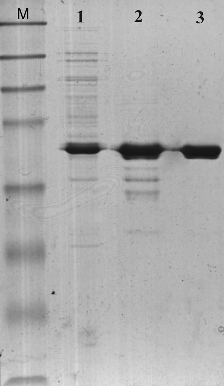
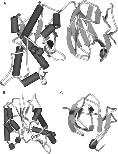
BH1115 is a gene from Bacillus halodurans strain C-125 that hypothetically encodes a rhamnogalacturonan acetyl esterase (RGAE) of the CE-12 family. As confirmation, this gene was cloned, and the product was expressed in Escherichia coli strain Rosetta (DE3) cells and purified. The enzyme obtained was monomeric, with a molecular mass of 45 kDa, and exhibited alkaliphilic properties. A study of the inhibition of the activity by some modulators confirmed that the catalytic triad for the esterase activity was Ser-His-Asp. This enzyme also presents broad substrate specificity and is active toward 7-aminocephalosporanic acid, cephalosporin C, p-nitrophenyl acetate, beta-naphthyl acetate, glucose pentaacetate, and acetylated xylan. Moreover, RGAE from B. halodurans achieves a synergistic effect with xylanase A toward acetylated xylan. As a member of the SGNH family, it does not adopt the common alpha/beta hydrolase fold. The homology between the folds of RGAE from Aspergillus aculeatus and the hypothetical YxiM precursor from Bacillus subtilis, which both belong to the SGNH family, illustrates the divergence of such proteins from a common ancestor. Furthermore, the enzyme possesses a putative substrate binding region at the N terminus of the protein which has never been described to date for any RGAE.
Figures

Similar articles
-
The SGNH hydrolase family: a template for carbohydrate diversity.Glycobiology. 2022 Sep 19;32(10):826-848. doi: 10.1093/glycob/cwac045. Glycobiology. 2022. PMID: 35871440 Free PMC article. Review.
-
YesT: a new rhamnogalacturonan acetyl esterase from Bacillus subtilis.Proteins. 2008 Apr;71(1):379-88. doi: 10.1002/prot.21705. Proteins. 2008. PMID: 17957779
-
A novel cephalosporin deacetylating acetyl xylan esterase from Bacillus subtilis with high activity toward cephalosporin C and 7-aminocephalosporanic acid.Appl Microbiol Biotechnol. 2014 Mar;98(5):2081-9. doi: 10.1007/s00253-013-5056-x. Epub 2013 Jul 5. Appl Microbiol Biotechnol. 2014. PMID: 23828600
-
Biochemical characterization of a GDSL-motif esterase from Bacillus sp. K91 with a new putative catalytic mechanism.J Microbiol Biotechnol. 2014 Nov 28;24(11):1551-8. doi: 10.4014/jmb.1406.06056. J Microbiol Biotechnol. 2014. PMID: 25394512
-
Rhamnogalacturonan acetylesterase elucidates the structure and function of a new family of hydrolases.Structure. 2000 Apr 15;8(4):373-83. doi: 10.1016/s0969-2126(00)00118-0. Structure. 2000. PMID: 10801485
Cited by
-
Characterization of a glucose-, xylose-, sucrose-, and D-galactose-stimulated β-glucosidase from the alkalophilic bacterium Bacillus halodurans C-125.Curr Microbiol. 2011 Mar;62(3):833-9. doi: 10.1007/s00284-010-9766-3. Epub 2010 Nov 3. Curr Microbiol. 2011. PMID: 21046402
-
Crystallization and diffraction analysis of Sm23: an SGNH-family arylesterase from Sinorhizobium meliloti 1021.Acta Crystallogr Sect F Struct Biol Cryst Commun. 2011 May 1;67(Pt 5):572-4. doi: 10.1107/S1744309111007706. Epub 2011 Apr 27. Acta Crystallogr Sect F Struct Biol Cryst Commun. 2011. PMID: 21543864 Free PMC article.
-
Enzymatic diversity of the Clostridium thermocellum cellulosome is crucial for the degradation of crystalline cellulose and plant biomass.Sci Rep. 2016 Oct 19;6:35709. doi: 10.1038/srep35709. Sci Rep. 2016. PMID: 27759119 Free PMC article.
-
Heterologous expression, purification and characterization of three novel esterases secreted by the lignocellulolytic fungus Penicillium purpurogenum when grown on sugar beet pulp.Carbohydr Res. 2017 Apr 18;443-444:42-48. doi: 10.1016/j.carres.2017.03.014. Epub 2017 Mar 18. Carbohydr Res. 2017. PMID: 28342968 Free PMC article.
-
The SGNH hydrolase family: a template for carbohydrate diversity.Glycobiology. 2022 Sep 19;32(10):826-848. doi: 10.1093/glycob/cwac045. Glycobiology. 2022. PMID: 35871440 Free PMC article. Review.
References
-
- Biely, P., J. Puls, and H. Schneider. 1985. Acetyl xylan esterases in fungal cellulolytic system. FEBS Lett. 18680-84.
-
- Choi, D. H., K. S. Han, I. S. Jeong, S. H. Lee, B. S. Lim, and Y. D. Kim. 1999. Cephalosporin deacetylase, gene coding for it, and preparation method of deacetylated cephalosporin compounds using it. WO patent (WO/1999/055881). Chem. Abstr. 131332993.
-
- Choi, D. H., Y. D. Kim, I. S. Chung, S. H. Lee, S. M. Kang, T. J. Kwon, and K. S. Han. 2000. Gene cloning and expression of cephalosporin-C deacetylase from Bacillus sp. KCCM10143. J. Microbiol. Biotechnol. 10221-226.
Publication types
MeSH terms
Substances
LinkOut - more resources
Full Text Sources
Molecular Biology Databases
Research Materials
