. 2008 Jul;190(13):4392-7.
doi: 10.1128/JB.01443-07. Epub 2007 Dec 14.

A mutational analysis defines Vibrio fischeri LuxR binding sites

Affiliations

A mutational analysis defines Vibrio fischeri LuxR binding sites

Luis Caetano M Antunes et al. J Bacteriol. 2008 Jul.

Abstract

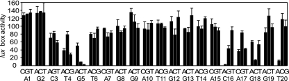
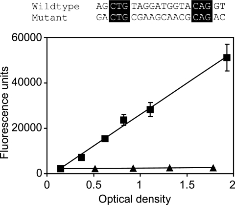
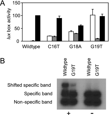
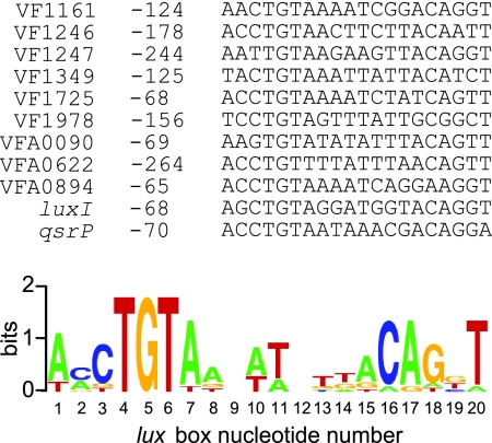
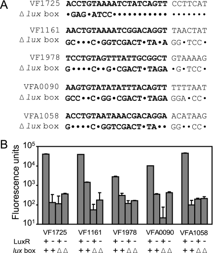
Vibrio fischeri quorum sensing involves the LuxI and LuxR proteins. The LuxI protein generates the quorum-sensing signal N-3-oxohexanoyl-l-homoserine lactone (3OC6-HSL), and LuxR is a signal-responsive transcriptional regulator which activates the luminescence (lux) genes and 17 other V. fischeri genes. For activation of the lux genes, LuxR binds to a 20-base-pair inverted repeat, the lux box, which is centered 42.5 base pairs upstream of the transcriptional start of the lux operon. Similar lux box-like elements have been identified in only a few of the LuxR-activated V. fischeri promoters. To better understand the DNA sequence elements required for LuxR binding and to identify binding sites in LuxR-regulated promoters other than the lux operon promoter, we have systematically mutagenized the lux box and evaluated the activity of many mutants. By doing so, we have identified nucleotides that are critical for promoter activity. Interestingly, certain lux box mutations allow a 3OC6-HSL-independent LuxR activation of the lux operon promoter. We have used the results of the mutational analysis to create a consensus lux box, and we have used this consensus sequence to identify LuxR binding sites in 3OC6-HSL-activated genes for which lux boxes could not be identified previously.


Figures

FIG. 1.
FIG. 1.
Effects of single-base-substitution lux box mutations on promoter activity. Every nucleotide in the lux box was replaced with the three other ones. The sequence of a wild-type lux box and the nucleotide numbering system used are shown on the bottom, and activity is expressed as percent of wild-type activity. Experiments were performed with recombinant E. coli cells containing the LuxR expression vector pHV402 and a plasmid containing a lux promoter-gfp transcriptional fusion with a lux box nucleotide substitution as indicated. Cultures were grown with 3OC6-HSL. The results represent the means of the results of three independent experiments, and the error bars represent standard deviations from the means.
FIG. 2.
FIG. 2.
Activity of a palindromic lux box containing substitutions in all nucleotides except those at positions 3 to 5 and 16 to 18. An alignment of the wild-type and mutant lux box sequences is shown. The unchanged nucleotides are boxed. Transcription was monitored in recombinant E. coli cells containing pHV402 and the appropriate lux promoter-gfp fusion plasmid. The activities of the wild-type lux box (squares) and the mutant lux box (triangles) are shown as a function of the optical density of the culture (at 600 nm). The results represent the means of the results of three independent experiments, and the error bars represent standard deviations.
FIG. 3.
FIG. 3.
3OC6-HSL-independent activity of lux box mutants. (A) In vivo transcriptional-fusion analysis of selected lux box mutants. Transcription was monitored as green fluorescent protein fluorescence in E. coli cells containing a lux promoter-gfp fusion plasmid with the indicated lux box. White bars are results for cells containing the LuxR expression vector pHV402 grown without 3OC6-HSL, gray bars are results for cells without pHV402 grown with 3OC6-HSL, and black bars are results for cells containing pHV402 grown with 3OC6-HSL. Data are presented as percent of the activity in cells with the wild-type lux box plasmid and pHV402 grown with 3OC6-HSL. The results are the means of the results of three independent experiments, and the error bars show standard deviations. (B) In vitro binding of purified LuxR to the wild-type lux box and the G19T mutant lux box with 3OC6-HSL (+) or without 3OC6-HSL (−). Specific probes were generated by PCR amplification of promoter regions using the transcriptional-fusion plasmids as templates. The nonspecific probe was obtained by PCR amplification of the multicloning site of the mini-CTX plasmid (15).
FIG. 4.
FIG. 4.
Predicted lux boxes found in the promoters of 3OC6-HSL-activated genes. The distances of the lux box elements from the translational start sites of the downstream open reading frames are indicated. The sequence logo shown was constructed based on the lux boxes found using WebLogo (http://weblogo.berkeley.edu/).
FIG. 5.
FIG. 5.
Mutational analysis of putative lux boxes of previously identified V. fisheri quorum-controlled promoters (1). (A) DNA sequences of putative lux boxes found in the promoter regions of the genes indicated and deletion mutants (Δlux box). Downstream DNA was identical in wild-type and mutant constructs. The lux box elements are in bold. Nucleotides unchanged in the mutant are shown as dots. (B) Promoter activity of constructs shown in panel A. Experiments were performed with recombinant E. coli cells, and data are given as fluorescence units (fluorescence per optical density unit). Activity in the presence (+) or absence (−) of LuxR is shown. Bars labeled “lux box +” represent the activity of wild-type constructs, whereas “lux box Δ” designates the activity of the Δlux box constructs shown in panel A. All fusions were tested in the presence of 3OC6-HSL. The results are the means of the results of three independent experiments. Error bars show standard deviations of the means.

Similar articles

Cited by

References

    1. Antunes, L. C. M., A. L. Schaefer, R. B. R. Ferreira, N. Qin, A. M. Stevens, E. G. Ruby, and E. P. Greenberg. 2007. A transcriptome analysis of the Vibrio fischeri LuxR-LuxI regulon. J. Bacteriol. 1898387-8391. - PMC - PubMed
    1. Bassler, B. L., and R. Losick. 2006. Bacterially speaking. Cell 125237-246. - PubMed
    1. Bassler, B. L., M. Wright, and M. R. Silverman. 1994. Multiple signalling systems controlling expression of luminescence in Vibrio harveyi: sequence and function of genes encoding a second sensory pathway. Mol. Microbiol. 13273-286. - PubMed
    1. Boettcher, K. J., and E. G. Ruby. 1990. Depressed light emission by symbiotic Vibrio fischeri of the sepiolid squid Euprymna scolopes. J. Bacteriol. 1723701-3706. - PMC - PubMed
    1. Devine, J. H., G. S. Shadel, and T. O. Baldwin. 1989. Identification of the operator of the lux regulon from the Vibrio fischeri strain ATCC7744. Proc. Natl. Acad. Sci. USA 865688-5692. - PMC - PubMed

Publication types

MeSH terms

LinkOut - more resources