. 2008 May;7(5):776-82.
doi: 10.1128/EC.00309-07. Epub 2007 Dec 14.

Candida albicans Als adhesins have conserved amyloid-forming sequences

Affiliations

Candida albicans Als adhesins have conserved amyloid-forming sequences

Henry N Otoo et al. Eukaryot Cell. 2008 May.

Abstract

The cell wall-bound Als adhesins of Candida albicans mediate both yeast-to-host tissue adherence and yeast aggregation. This aggregation is amyloid-like, with self-propagating secondary-structure changes, amyloid-characteristic dye binding, and induced birefringence (J. M. Rauceo, N. K. Gaur, K. G. Lee, J. E. Edwards, S. A. Klotz, and P. N. Lipke, Infect. Immun. 72:4948-4955, 2004). Therefore, we determined whether Als proteins could form amyloid fibers with properties like those in cellular aggregation. The beta-aggregation predictor TANGO identified a heptapeptide sequence present in a highly conserved sequence with amyloid-forming potential in Als1p, Als3p, and Als5p. A tridecapeptide containing this sequence formed fibers that bound Congo red and thioflavin T and had characteristic amyloid morphology. Als5p(20-431) and Als5p(20-664), large fragments of Als5p containing the amyloid sequence, also formed amyloid-like fibers and bound Congo red under native conditions. K(a)/K(s) analysis showed that the amyloid-forming sequences are highly conserved in Als proteins and evolve more slowly than other regions of the proteins. Therefore, amyloid-forming ability itself is conserved in these proteins.


Figures
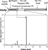
FIG. 1.
FIG. 1.
(Top) Map of regions of Als5p. Specific domains are labeled. (Bottom) TANGO β-aggregation potential for the soluble fragment Als5p20-620. The position of the major peak is marked on the protein map.
FIG. 2.
FIG. 2.
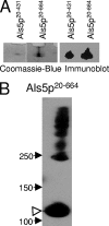
Gel electrophoresis of Als5p. All gels were 4 to 20% acrylamide gradient gels. (A) SDS gel of partially solubilized precipitates from preparations of purified Als5p fragments stained with Coomassie blue (left) or immunoblotted with anti-V5 (right). The precipitates were isolated and washed three times before electrophoresis. The illustrated region of the gel was immediately below the loading wells. (B) Immunoblot of Als5p20-664 showing multimeric aggregates in the presence of SDS. The protein was concentrated 10-fold after purification, and an aliquot of the suspension was treated with SDS and then electrophoresed for 24 h. The filled arrowheads mark the positions of Bio-Rad Rainbow molecular weight markers. The open arrowhead marks the Als5p20-664 monomer with an apparent molecular mass of 120 kDa (24).
FIG. 3.
FIG. 3.
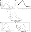
Absorbance and fluorescence spectra. (A) Congo red absorbance spectrum (solid line) and spectrum of Congo red in the presence of the unstirred “unaggregated” peptide S322NGIVIVATTRTV334 (dotted line) or the aggregated peptide S322NGIVIVATTRTV334 (dashed line). The peptide concentration was 20 μg/ml. (B) Congo red absorbance spectra. The curve with maximal absorbance is in the absence of added peptide. Adding the “mutant” peptide SNGINIVATTRTV that had been incubated with or without stirring slightly diluted the dye but did not otherwise change the spectrum (superimposed spectra with slightly lower absorbance maxima). The peptide concentration was 20 μg/ml. (C) Fluorescence emission spectra for thioflavin T (solid line), thioflavin T in the presence of an unstirred “unaggregated” peptide (dotted line), and thioflavin T in the presence of aggregated peptide (dashed line). The peptide concentration was 6.6 μg/ml. (D) Congo red difference spectra for unaggregated (solid line) or aggregated (dashed line) Als5p20-431. Absorbance values for Congo red alone have been subtracted (maximal absorbance, 0.0510 at 489 nm). The final concentration of protein was 5 μg/ml. (E) Congo red difference spectra for unaggregated (solid line) or aggregated (dotted line) Als5p20-664. Absorbance values for Congo red alone have been subtracted (maximal absorbance, 0.0646 at 502 nm). The final concentration of protein was 3.5 μg/ml.
FIG. 4.
FIG. 4.
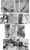
Gallery of fibers. (A) fibers formed by tridecamer peptide; the arrowheads in A3 show helical striations. Bar lengths: A1, 500 nm; A2 and A3, 100 nm. (B) Als5p20-431. (B1) Nonfibrous aggregate; the bar length is 200 nm. (B2 to B4) Fibers with bar lengths of 500 nm. (C) Fibers formed from Als5p20-664. Bar lengths: C1 to C3, 500 nm; C4, 2 μm.

References

    1. Barnhart, M. M., and M. R. Chapman. 2006. Curli biogenesis and function. Annu. Rev. Microbiol. 60131-147. - PMC - PubMed
    1. Chandra, J., D. M. Kuhn, P. K. Mukherjee, L. L. Hoyer, T. McCormick, and M. A. Ghannoum. 2001. Biofilm formation by the fungal pathogen Candida albicans: development, architecture, and drug resistance. J. Bacteriol. 1835385-5394. - PMC - PubMed
    1. Chapman, M. R., L. S. Robinson, J. S. Pinkner, R. Roth, J. Heuser, M. Hammar, S. Normark, and S. J. Hultgren. 2002. Role of Escherichia coli curli operons in directing amyloid fiber formation. Science 295851-855. - PMC - PubMed
    1. Chimpanzee Sequencing and Analysis Consortium. 2005. Initial sequence of the chimpanzee genome and comparison with the human genome. Nature 43769-87. - PubMed
    1. Dranginis, A. M., J. R. Rauceo, J. E. Coronado, and P. N. Lipke. 2007. A biochemical guide to yeast adhesins: glycoproteins for social and antisocial occasions. Microbiol. Mol. Biol. Rev. 71282-294. - PMC - PubMed

Publication types

MeSH terms

LinkOut - more resources