

Proteasomal degradation of human release factor eRF3a regulates translation termination complex formation
- PMID: 18083835
- PMCID: PMC2212242
- DOI: 10.1261/rna.728608
Proteasomal degradation of human release factor eRF3a regulates translation termination complex formation
Abstract
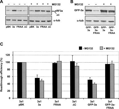
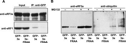
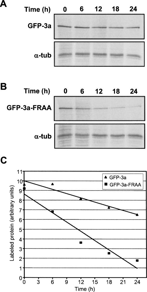
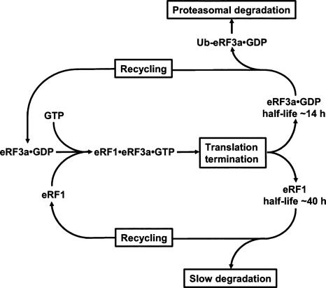
In eukaryotes, eRF1 and eRF3 are associated in a complex that mediates translation termination. The regulation of the formation of this complex in vivo is far from being understood. In mammalian cells, depletion of eRF3a causes a reduction of eRF1 level by decreasing its stability. Here, we investigate the status of eRF3a when not associated with eRF1. We show that eRF3a forms altered in their eRF1-binding site have a decreased stability, which increases upon cell treatment with the proteasome inhibitor MG132. We also show that eRF3a forms altered in eRF1 binding as well as wild-type eRF3a are polyubiquitinated. These results indicate that eRF3a is degraded by the proteasome when not associated with eRF1 and suggest that proteasomal degradation of eRF3a controls translation termination complex formation by adjusting the eRF3a level to that of eRF1.
Figures
Similar articles
-
Involvement of human release factors eRF3a and eRF3b in translation termination and regulation of the termination complex formation.Mol Cell Biol. 2005 Jul;25(14):5801-11. doi: 10.1128/MCB.25.14.5801-5811.2005. Mol Cell Biol. 2005. PMID: 15987998 Free PMC article.
-
PABP enhances release factor recruitment and stop codon recognition during translation termination.Nucleic Acids Res. 2016 Sep 19;44(16):7766-76. doi: 10.1093/nar/gkw635. Epub 2016 Jul 14. Nucleic Acids Res. 2016. PMID: 27418677 Free PMC article.
-
Human eukaryotic release factor 3a depletion causes cell cycle arrest at G1 phase through inhibition of the mTOR pathway.Mol Cell Biol. 2007 Aug;27(16):5619-29. doi: 10.1128/MCB.00035-07. Epub 2007 Jun 11. Mol Cell Biol. 2007. PMID: 17562865 Free PMC article.
-
Termination of translation in eukaryotes.Biochem Cell Biol. 1995 Nov-Dec;73(11-12):1079-86. doi: 10.1139/o95-116. Biochem Cell Biol. 1995. PMID: 8722024 Review.
-
Translation termination and its regulation in eukaryotes: recent insights provided by studies in yeast.Biochemistry (Mosc). 1999 Dec;64(12):1360-6. Biochemistry (Mosc). 1999. PMID: 10648959 Review.
Cited by
-
Studies on human eRF3-PABP interaction reveal the influence of eRF3a N-terminal glycin repeat on eRF3-PABP binding affinity and the lower affinity of eRF3a 12-GGC allele involved in cancer susceptibility.RNA Biol. 2016;13(3):306-15. doi: 10.1080/15476286.2015.1137421. Epub 2016 Jan 28. RNA Biol. 2016. PMID: 26818177 Free PMC article.
-
Phosphorylation of elongation factor-2 kinase differentially regulates the enzyme's stability under stress conditions.Biochem Biophys Res Commun. 2012 Jul 27;424(2):308-14. doi: 10.1016/j.bbrc.2012.06.112. Epub 2012 Jun 27. Biochem Biophys Res Commun. 2012. PMID: 22749997 Free PMC article.
-
Mammalian Hbs1L deficiency causes congenital anomalies and developmental delay associated with Pelota depletion and 80S monosome accumulation.PLoS Genet. 2019 Feb 1;15(2):e1007917. doi: 10.1371/journal.pgen.1007917. eCollection 2019 Feb. PLoS Genet. 2019. PMID: 30707697 Free PMC article.
-
A Computational Recognition Analysis of Promising Prognostic Biomarkers in Breast, Colon and Lung Cancer Patients.Int J Mol Sci. 2025 Jan 25;26(3):1017. doi: 10.3390/ijms26031017. Int J Mol Sci. 2025. PMID: 39940786 Free PMC article.
-
Genetic association and functional characterization of MCPH1 gene variation in bipolar disorder and schizophrenia.Am J Med Genet B Neuropsychiatr Genet. 2019 Jun;180(4):258-265. doi: 10.1002/ajmg.b.32722. Epub 2019 Mar 11. Am J Med Genet B Neuropsychiatr Genet. 2019. PMID: 30859703 Free PMC article.
References
-
- Alkalaeva, E.Z., Pisarev, A.V., Frolova, L.Y., Kisselev, L.L., Pestova, T.V. In vitro reconstitution of eukaryotic translation reveals cooperativity between release factors eRF1 and eRF3. Cell. 2006;125:1125–1136. - PubMed
-
- Chabelskaya, S., Kiktev, D., Inge-Vechtomov, S., Philippe, M., Zhouravleva, G. Nonsense mutations in the essential gene SUP35 of Saccharomyces cerevisiae are nonlethal. Mol. Genet. Genomics. 2004;272:297–307. - PubMed
-
- Corish, P., Tyler-Smith, C. Attenuation of green fluorescent protein half-life in mammalian cells. Protein Eng. 1999;12:1035–1040. - PubMed
-
- Dantuma, N.P., Lindsten, K., Glas, R., Jellne, M., Masucci, M.G. Short-lived green fluorescent proteins for quantifying ubiquitin/proteasome-dependent proteolysis in living cells. Nat. Biotechnol. 2000;18:538–543. - PubMed
Publication types
MeSH terms
Substances
LinkOut - more resources
Full Text Sources