. 2008 Apr 1;111(7):3313-21.
doi: 10.1182/blood-2007-08-110114. Epub 2007 Dec 14.

Rac1 is essential for intraembryonic hematopoiesis and for the initial seeding of fetal liver with definitive hematopoietic progenitor cells

Affiliations

Rac1 is essential for intraembryonic hematopoiesis and for the initial seeding of fetal liver with definitive hematopoietic progenitor cells

Gabriel Ghiaur et al. Blood. .

Abstract

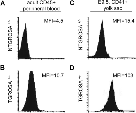
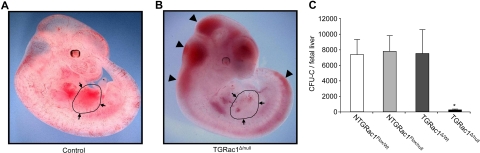
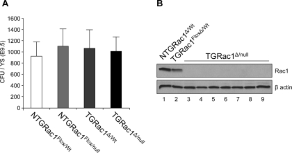
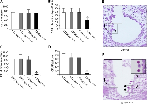
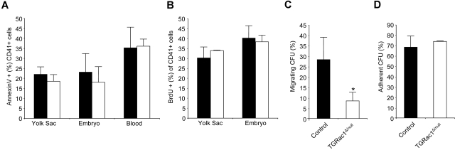
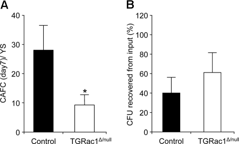
Definitive hematopoietic stem and progenitor cells (HSCs/Ps) originating from the yolk sac and/or para-aorta-splanchno-pleura/aorta-gonad-mesonephros are hypothesized to colonize the fetal liver, but mechanisms involved are poorly defined. The Rac subfamily of Rho GTPases has been shown to play essential roles in HSC/P localization to the bone marrow following transplantation. Here, we study the role of Rac1 in HSC/P migration during ontogeny and seeding of fetal liver. Using a triple-transgenic approach, we have deleted Rac1 in HSCs/Ps during very early embryonic development. Without Rac1, there was a decrease in circulating HSCs/Ps in the blood of embryonic day (E) 10.5 embryos, while yolk sac definitive hematopoiesis was quantitatively normal. Intraembryonic hematopoiesis was significantly impaired in Rac1-deficient embryos, culminating with absence of intra-aortic clusters and fetal liver hematopoiesis. At E10.5, Rac1-deficient HSCs/Ps displayed decreased transwell migration and impaired inter-action with the microenvironment in migration-dependent assays. These data suggest that Rac1 plays an important role in HSC/P migration during embryonic development and is essential for the emergence of intraembryonic hematopoiesis.


Figures

Figure 1
Figure 1
Expression profile of Vav1-driven Cre-mediated recombination. Flow cytometric analysis of recombination status of RosaGFP locus in CD41+ definitive hematopoietic progenitors from E9.5 yolk sac (right column) and CD45+ adult peripheral blood leukocytes (left column). As a negative control, a Vav1Cre nontransgenic (NTG) ROSA heterozygous mouse is presented in the top row. The mean fluorescence intensity (MFI) of the GFP channel is depicted in each panel. Data are representative of 3 independent specimens analyzed for each genotype (N = 3).
Figure 2
Figure 2
Phenotypic analysis of embryos at E11.5. (A,B) Gross appearance of surviving TGRac1Flox/null embryos and littermate controls at E11.5. The limbs from the right side of the embryos were removed for clarity of presentation. The appearance of the littermate controls was similar for NTGRac1Flox/Wt, NTGRac1Flox/null, and TGRac1Flox/Wt (hereafter referred to as “control”). The anatomic region of the fetal liver is delineated and noted by arrows. The arrowheads point to accumulation of blood in cephalic regions as well as areas underlying the spinal cord, a phenotype previously noted in mutant animals deficient in hematopoietic cell production at the fetal liver stage. (C) Quantitative analysis of fetal liver hematopoiesis as analyzed by CFU/FL of F2 embryos at E11.5. Data represent mean (± SD) of at least 3 embryos analyzed for each genotype. *P < .01, TGRac1Flox/null versus all other genotypes.
Figure 3
Figure 3
Definitive hematopoiesis in the yolk sac at E9.5. (A) Quantitative analysis of definitive hematopoietic progenitor cells as analyzed by CFU/YS of F2 embryos at E9.5. Data represent mean (± SD) of at least 7 embryos analyzed for each genotype. There was no significant difference among genotypes. (B) Immunoblot analysis of Rac1 protein expression level of individual hematopoietic progenitor colonies from the yolk sac of TGRac1Flox/null mice (lanes 3-9) and control mice (lanes 1-2) at E9.5. β-Actin is shown as a loading control. Note intensity of Rac1 band in lane 2 versus lane 1, representing expression of Rac1 in the heterozygous versus WT genotype. These results are representative of 3 independent experiments with similar results.
Figure 4
Figure 4
Definitive hematopoiesis of embryos at E10.5. (A-D) Quantitative analysis of definitive hematopoiesis at E10.5 by determination of number of CFUs in (A) yolk sac; (B) blood; (C) embryonic remnants (embryonic tissue after removal of yolk sac and amniotic membranes, blood, fetal liver, and tail bud, used for genotyping); and (D) fetal liver. Data represent means (± SD) of 8 or more F2 embryos for each genotype, *P < .01, TGRac1Flox/null versus all other genotypes. (E,F) Histologic analysis of the aortic clusters at E10.5. Punctuated squares delineate the area shown at higher magnification in the upper-right corner. The figures are representative of all animals analyzed for each genotype. Data are summated in Table 2. Arrow in panel E shows hematopoietic cells on the ventral wall of dorsal aorta. Arrowheads in panel F show circulating primitive red blood cells. No TGRac1Flox/null mice demonstrated aortic clusters. Images acquired as described in “Light microscopy,” with a 40×/0.5 numeric aperture objective.
Figure 5
Figure 5
Apoptosis, proliferation, adhesion, and migration of definitive hematopoietic progenitors at E10.5. (A) Level of apoptosis of CD41+ definitive hematopoietic progenitors from different embryonic tissues at E10.5 analyzed by annexinV/7AAD staining and flow cytometry. Figure shows one representative experiment of 3 with similar results. Data represent means (± SD) of at least 3 embryos analyzed for each genotype. Differences between genotypes were not significant. (B) BrdU incorporation of definitive hematopoietic progenitors isolated from the yolk sac or the embryo proper of F2 embryos at E10.5. Figure shows one representative experiment of 3 with similar results. Data represent means (± SD) of at least 3 embryos analyzed for each genotype. Differences among genotypes were not significant. (C) SCF/SDF1-α induced directed migration on FN CH296-coated transwell filters of circulating E10.5 definitive hematopoietic progenitors. Data represent means (± SD) of 3 independent experiments. (D) β1-integrin–mediated adhesion to FN CH296 of circulating E10.5 definitive hematopoietic progenitors assayed by CFU. Data represent means (± SD) of 3 independent experiments. There was no significant difference among groups. *P < .05, TGRac1Flox/null versus control (N = 3). (■ indicates littermate control; □, TGRac1Flox/null).
Figure 6
Figure 6
In vitro characterization of interaction between yolk sac–derived E10.5 definitive hematopoietic progenitors and stromal cells. (A) Total number of CAFCs per yolk sac of E10.5 F2 embryos. Data represent means (± SD) of 3 independent experiments. *P < .05, TGRac1Flox/null versus littermate controls (N = 3). (B) Survival of definitive hematopoietic progenitors from the yolk sac at E10.5 in a hanging drop organ culture with AFT024 stroma cells (“Methods”). Data represent means (± SD) of 3 independent experiments (N = 3). There was no significant difference between genotypes. ■ indicates littermate control; □, TGRac1Flox/null.

References

    1. Gekas C, Dieterlen-Lievre F, Orkin SH, Mikkola HK. The placenta is a niche for hematopoietic stem cells. Dev Cell. 2005;8:365–375. - PubMed
    1. Moore MA, Metcalf D. Ontogeny of the haemopoietic system: yolk sac origin of in vivo and in vitro colony forming cells in the developing mouse embryo. Br J Hematol. 1970;18:279–296. - PubMed
    1. Weissman I, Papaioannou V, Gardner R. Fetal hematopoietic origins of the adult hematolymphoid system. In: Clarkson B, Marks PA, Till JE, editors. Differentiation of Normal and Neoplastic Hematopoietic Cells. Cold Spring Harbor, NY: Cold Spring Harbor Laboratory; 1978. pp. 371–387.
    1. Johnson GR, Moore MA. Role of stem cell migration in initiation of mouse foetal liver haemopoiesis. Nature. 1975;258:726–728. - PubMed
    1. Palis J, Chan RJ, Koniski A, Patel R, Starr M, Yoder MC. Spatial and temporal emergence of high proliferative potential hematopoietic precursors during murine embryogenesis. Proc Natl Acad Sci U S A. 2001;98:4528–4533. - PMC - PubMed

Publication types

MeSH terms