. 2007 Nov:1116:82-99.
doi: 10.1196/annals.1402.082.

Axin1 and Axin2 are regulated by TGF- and mediate cross-talk between TGF- and Wnt signaling pathways

Affiliations

Axin1 and Axin2 are regulated by TGF- and mediate cross-talk between TGF- and Wnt signaling pathways

Debbie Y Dao et al. Ann N Y Acad Sci. 2007 Nov.

Abstract

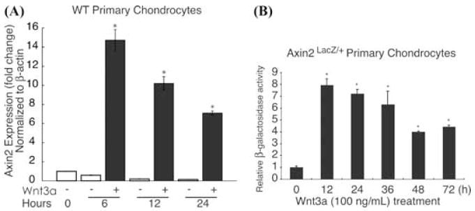
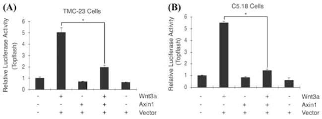
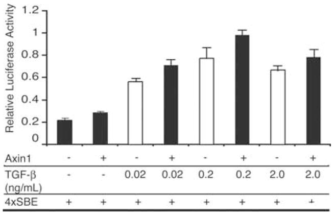
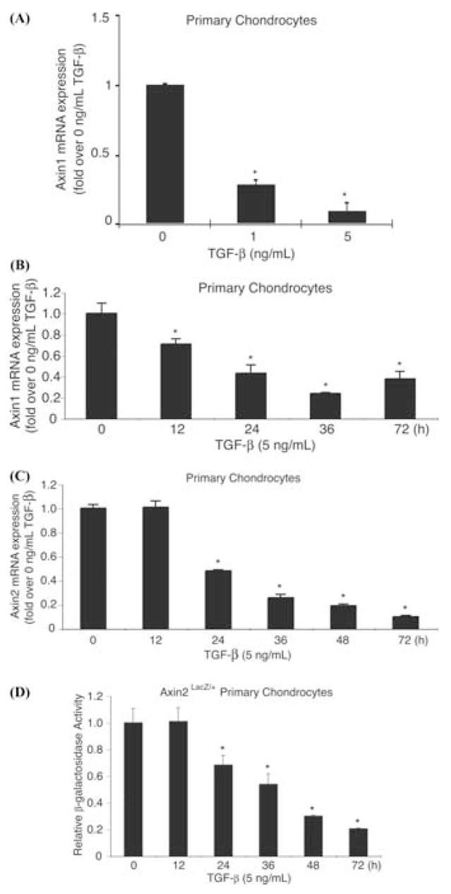
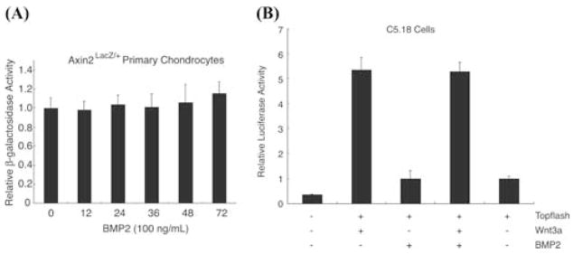
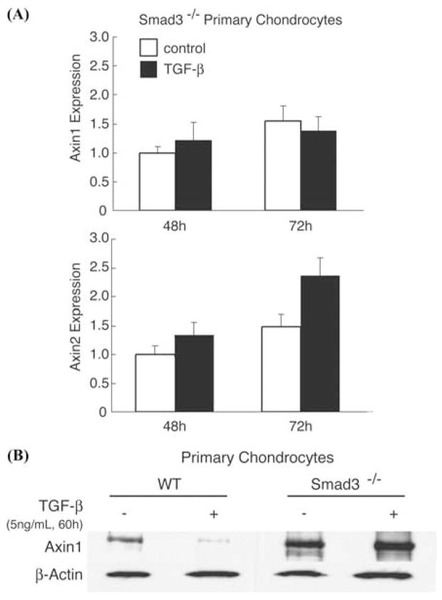
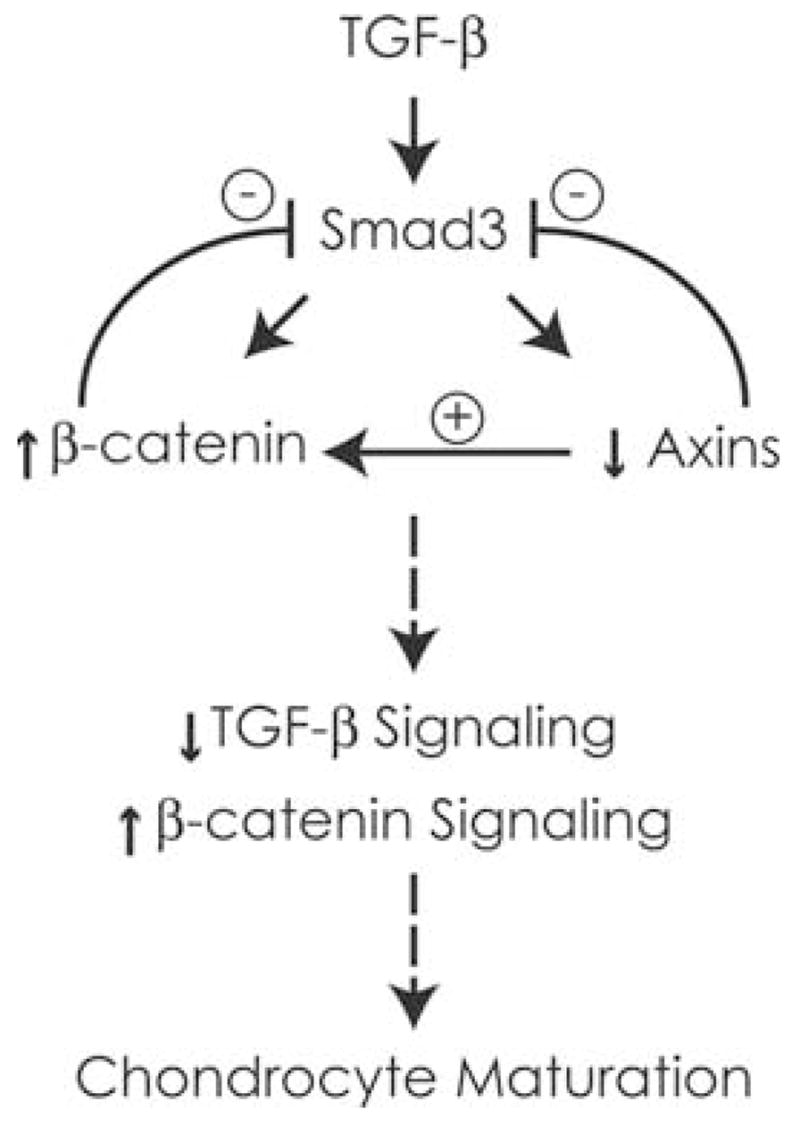
Chondrocyte maturation during endochondral bone formation is regulated by a number of signals that either promote or inhibit maturation. Among these, two well-studied signaling pathways play crucial roles in modulating chondrocyte maturation: transforming growth factor-beta (TGF-beta)/Smad3 signaling slows the rate of chondrocyte maturation, while Wingless/INT-1-related (Wnt)/beta-catenin signaling enhances the rate of chondrocyte maturation. Axin1 and Axin2 are functionally equivalent and have been shown to inhibit Wnt/beta-catenin signaling and stimulate TGF-beta signaling. Here we show that while Wnt3a stimulates Axin2 in a negative feedback loop, TGF-beta suppresses the expression of both Axin1 and Axin2 and stimulates beta-catenin signaling. In Axin2 -/- chondrocytes, TGF-beta treatment results in a sustained increase in beta-catenin levels compared to wild-type chondrocytes. In contrast, overexpression of Axin enhanced TGF-beta signaling while overexpression of beta-catenin inhibited the ability of TGF-beta to induce Smad3-sensitive reporters. Finally, the suppression of the Axins is Smad3-dependent since the effect is absent in Smad3 -/- chondrocytes. Altogether these findings show that the Axins act to integrate signals between the Wnt/beta-catenin and TGF-beta/Smad pathways. Since the suppression Axin1 and Axin2 expression by TGF-beta reduces TGF-beta signaling and enhances Wnt/beta-catenin signaling, the overall effect is a shift from TGF-beta toward Wnt/beta-catenin signaling and an acceleration of chondrocyte maturation.


Figures

FIGURE 1
FIGURE 1
Wnt3a induces Axin2 gene and protein expression. Primary murine sternal chondrocytes were isolated and placed in monolayer culture as described in “Methods.” After overnight culture they were treated with Wnt3a (100 ng/mL) or with a vehicle control for 0, 6, 12, and 24 h before total RNA was isolated. Quantitative Reverse-Transcriptase PCR was performed to assess Axin2 gene expression (normalized to β-actin) (A). Primary chondrocytes from Axin2LacZ/+ mice were placed in monolayer culture overnight and then treated with 100 ng/mL of Wnt3a. Cultures were harvested after 0, 12, 24, 36, 48, and 72 h and and β-galactosidase activity was measured as a reporter of Axin2 protein expression (B). The symbol * represents significance at P < 0.05 when compared to 0 h.
FIGURE 2
FIGURE 2
Axin inhibits Wnt3a signaling in chondrocytes. TMC23 (A) and C5.18 (B) chondrocyte cell lines were transfected with the Topflash reporter construct and treated with Wnt3a (100 ng/mL) in the presence or absence of cotransfection with Axin1. Cell extracts were harvested after 48 h and luciferase activity measured. Transfection efficiency was determined by measurement of renilla luciferase activity. The symbol * represents significance at P < 0.05.
FIGURE 3
FIGURE 3
β-catenin inhibits TGF-β signaling in chondrocytes while the induction of β-catenin by TGF-β is enhanced in Axin2-deficient chondrocytes. (A) Chondrocyte C5.18 cells were transfected with TGF-β responsive reporter, p3TP-Lux in the presence or absence of cotransfections with Smad3 and/or β-catenin and treated with or without TGF-β (5 ng/mL) for 48 h. Transfection of β-catenin significantly inhibited TGF-β or TGF-β+Smad3-induced p3TP-Lux reporter activity (*P < 0.05, **P < 0.01). (B) Protein lysates from TGF-β-treated primary chondrocytes from WT and Axin2−/− mice were assayed for β-catenin by Western blot. TGF-β treatment enhances β-catenin accumulation in both WT and Axin2−/− cells. However, the elevated levels of β-catenin persist in Axin2 −/− cells.
FIGURE 4
FIGURE 4
Axin enhances TGF-β signaling in chondrocytes. C5.18 chondrocyte cultures were transfected with 4×SBE and treated with 0.02, 0.2, or 2.0 ng/mL of TGF-β in the presence of absence of cotransfection with Axin plasmid. Cultures were harvested after 24 h and show that Axin cotransfection consistently enhanced reporter activity approximately 20% at each TGF-β concentration.
FIGURE 5
FIGURE 5
TGF-β inhibits Axin1 and Axin2 expression in chondrocytes. Primary murine chondrocytes were isolated and placed in overnight culture. The cultures were then treated with TGF-β (1 or 5 ng/mL) and total RNA was harvested at 48 h. Real-time RT PCR was used to determine the effect on Axin1 gene expression (A). Chondrocyte cultures with treated with TGF-β (5 ng/mL) and total RNA harvested at various times between 0 and 72 h and real-time RT-PCR used to determine the effect on expressions of Axin1 (B) and Axin2 (C) gene expression. β-galactosidase activity was measured in primary chondrocytes from Axin2LacZ/+ knockin mice and revealed that Axin2 protein expression is also negatively regulated by TGF-β progressively over time (D). The symbol *represents significance at P < 0.05 when compared to 0 ng/mL TGF-β or 0 h.
FIGURE 6
FIGURE 6
BMP does not alter Axin expression or Wnt/β-catenin signaling. Primary chondrocyte cells from Axin2LacZ/+ knockin mice were treated with BMP-2 (100 ng/mL) and cell extracts harvested between 0 and 72 h. β-galactosidase reporter activity was measured (A). C5.18 cells were transfected with the Wnt Topflash reporter and treated with BMP-2 (100 ng/mL) in the presence or absence of Wnt3a. Cell extracts were harvested after 24 h and luciferase activity was measured. The luciferase activity was normalized to renilla luciferase (B).
FIGURE 7
FIGURE 7
Smad3 −/− cells do not exhibit TGF-β-mediated inhibition of Axin expression. Primary chondrocytes from Smad 3−/− mice were isolated and cultured with 5 ng/mL TGF-β treatment for 24 and 48 h. Total RNA was harvested and RT-PCR performed to measure the relative suppression of Axin1 or Axin2 gene expression in TGF-β-treated cultures compared to control cultures after 48 h of treatment (A). Total cell protein extracts were obtained from wild-type and Smad3 −/− chondrocyte cultures treated with TGF-β or control medium and Axin1 protein levels measured by Western blot. β-actin was used as a loading control (B).
FIGURE 8
FIGURE 8
Summary of TGF-β/Smad and β-catenin signaling interactions in chondrocytes. TGF-β signaling inhibits chondrocyte maturation and induces Wnt/β-catenin signaling by directly activating β-catenin and reducing expression of Axin1 and Axin2. While β-catenin is necessary for the induction of cyclin D1 and chondrocyte proliferation downstream of TGF-β/Smad3, the induction of β-catenin and suppression of axins result in a decrease in TGF-β signaling. The overall effect of enhanced β-catenin signaling and inhibited Smad3 signaling is a release of chondrocytes from the inhibitory effects of TGF-β signaling on maturation. β-catenin in the presence of other stimulators of maturation, including BMP signaling molecules and Runx2, enhances chondrocyte maturation.

Similar articles

Cited by

References

    1. Ferguson CM, et al. Smad 2 and 3 mediate TGF-β1-induced inhibition of chondrocyte maturation. Endocrinology. 2000;141:4728–4735. - PubMed
    1. Li TF, et al. Smad3-deficient chondrocytes have enhanced BMP signaling and accelerated differentiation. J Bone Miner Res. 2006;21:4–16. - PMC - PubMed
    1. Kobayashi T, et al. BMP signaling stimulates cellular differentiation at multiple steps during cartilage development. Proc Natl Acad Sci USA. 2005;102:18023–18027. - PMC - PubMed
    1. Iwamoto M, et al. Runx2 expression and action in chondrocytes are regulated by retinoid signaling and parathyroid hormone-related peptide (PTHrP) Osteoarthr Cart. 2003;11:6–15. - PubMed
    1. Ionescu AM, et al. PTHrP modulates chondrocyte differentiation through AP-1 and CREB signaling. J Biol Chem. 2001;276:11639–11647. - PubMed

Publication types