. 2008 Feb;36(3):839-48.
doi: 10.1093/nar/gkm874. Epub 2007 Dec 15.

Analysis of intermolecular base pair formation of prohead RNA of the phage phi29 DNA packaging motor using NMR spectroscopy

Affiliations

Analysis of intermolecular base pair formation of prohead RNA of the phage phi29 DNA packaging motor using NMR spectroscopy

Aya Kitamura et al. Nucleic Acids Res. 2008 Feb.

Abstract

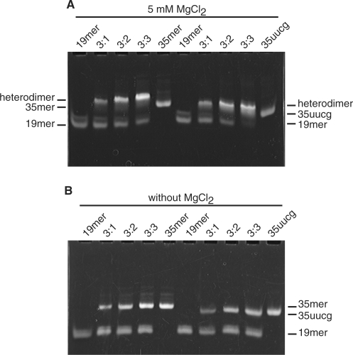
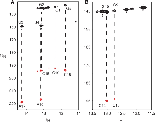
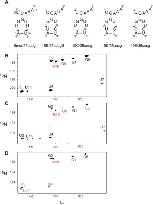
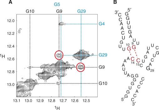
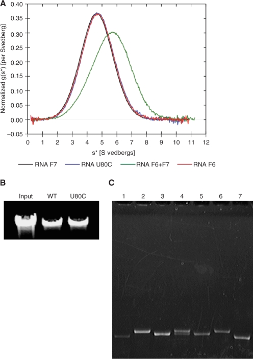
The bacteriophage ø29 DNA packaging motor that assembles on the precursor capsid (prohead) contains an essential 174-nt structural RNA (pRNA) that forms multimers. To determine the structural features of the CE- and D-loops believed to be involved in multimerization of pRNA, 35- and 19-nt RNA molecules containing the CE-loop or the D-loop, respectively, were produced and shown to form a heterodimer in a Mg2+-dependent manner, similar to that with full-length pRNA. It has been hypothesized that four intermolecular base pairs are formed between pRNA molecules. Our NMR study of the heterodimer, for the first time, proved directly the existence of two intermolecular Watson-Crick G-C base pairs. The two potential intermolecular A-U base pairs were not observed. In addition, flexibility of the D-loop was found to be important since a Watson-Crick base pair introduced at the base of the D-loop disrupted the formation of the intermolecular G-C hydrogen bonds, and therefore affected heterodimerization. Introduction of this mutation into the biologically active 120-nt pRNA (U80C mutant) resulted in no detectable dimerization at ambient temperature as shown by native gel and sedimentation velocity analyses. Interestingly, this pRNA bound to prohead and packaged DNA as well as the wild-type 120-nt pRNA.


Figures

Figure 1.
Figure 1.
Predicted secondary structures of ø29 pRNA and model RNAs. (A) The 120-nt form of wild-type pRNA. The shaded residues in the CE-loop and the D-loop are considered to form intermolecular base pairs. (B) The 19-mer that contains the D-loop and the D-helix. The G–U base pair at the end of the D-helix was changed to G–C, and an additional G–C base pair added in order to stabilize the helix. (C) The 35-mer that contains a part of the C-helix, the CE-loop, the E-helix and the E-loop. A G–C base pair was added at the end of the C-helix in order to stabilize the helix, and the U-bulge in the C-helix has been removed. (D) 35 -uucg in which the UUCG tetraloop (colored in red) is substituted for the E-loop (bases 20–25).
Figure 2.
Figure 2.
Non-denaturing PAGE analysis of RNAs (A) with 5 mM MgCl2 and (B) without MgCl2. The 19-mer was mixed with varying ratios of 35-mer or 35 -uucg and heterodimer formation was assessed by native gel electrophoresis. The lanes in each gel contain: (1) 0.45 nmol 19-mer; (2) 0.45 nmol 19-mer and 0.15 nmol 35-mer; (3) 0.45 nmol 19-mer and 0.30 nmol 35-mer; (4) 0.45 nmol 19-mer and 0.45 nmol 35-mer and (5) 0.45 nmol 35-mer. In lanes (6–10), 35 -uucg was used instead of 35-mer.
Figure 3.
Figure 3.
Sequential assignment of the 19-mer/35 -uucg heterodimer. H6/H8-H1′ region of the 2D-NOESY spectrum of the 19-mer/35 -uucg heterodimer is shown. The red solid lines indicate the sequential NOEs from G1 to G6 and from C31 to C34 in 35 -uucg. The red dashed lines indicate the sequential NOEs from C16 to U21 and from C27 to A29 in 35 -uucg. The blue solid lines indicate the NOEs from G1 to A6 in the 19-mer. The blue dashed lines indicate the NOEs from C15 to C19 in the 19-mer.
Figure 4.
Figure 4.
Identification of intermolecular base pairs. (A) HNN-COSY spectrum of the 15N-labeled 19-mer/non-labeled 35 -uucg heterodimer, in which only intramolecular hydrogen bonds in the 19-mer are observed. Lines indicate the intramolecular correlation between imino groups and acceptor nitrogens. (B) HNN-COSY spectrum of the G-labeled 19-mer/C-labeled 35 -uucg heterodimer, in which hydrogen bonds within the intermolecular G–C base pairs are observed.
Figure 5.
Figure 5.
19-mer variants. (A) Sequence of the RNA variants of the D-loop of the 19-mer and the CE-loop of 35 -uucg. Substituted residues are shown in red. (B-D) Imino region of 1H-15N HSQC spectra of (B) the labeled 19-mer/non-labeled 35 -uucg heterodimer, of (C) the labeled 19C/non-labeled 35 -uucg mixture and of (D) the labeled 19E/non-labeled 35 -uucg mixture.
Figure 6.
Figure 6.
Intermolecular NOE between G29 and G9. (A) Imino–imino region of 2D-NOESY of the 19-mer/35-uucg. Dashed black lines indicate imino proton resonances derived from the 19-mer. Dashed blue lines indicate resonances derived from 35 -uucg. NOEs between G29 of 35 -uucg and G9 in the 19-mer are indicated by red circles. (B) Schematic representation of the intermolecular base pairs between the 19-mer and 35 -uucg.
Figure 7.
Figure 7.
Dimerization analysis of the E variant 19-mer. The 19-mer or the E variant (19E) was mixed with varying ratios of 35 -uucg and heterodimer formation assessed by native gel electrophoresis. The lanes contain: (1) 0.45 nmol 19-mer; (2) 0.45 nmol 19-mer and 0.15 nmol 35 -uucg; (3) 0.45 nmol 19-mer and 0.30 nmol 35 -uucg; (4) 0.45 nmol 19-mer and 0.45 nmol 35 -uucg. In lanes (5–8), 19E was used instead of 19-mer. (9) 0.45 nmol 35 -uucg.
Figure 8.
Figure 8.
Analysis of the U80C mutant. (A) Ultracentrifugation sedimentation velocity analysis. Overlay of the normalized g(s*) plots from DcDt+ analysis (37,38). F6 and F7 are monomeric 120-nt pRNA variants that form a dimer only when they are mixed (5). The F6 + F7 sample appears to be a mixture of 48% monomer and 52% dimer (Supplementary Figure S4). (B) DNA packaging assay. DNA packaged by proheads with pRNA or proheads reconstituted with U80C was extracted and analyzed by agarose gel electrophoresis. The negative control (no ATP) showed no packaging (data not shown). (C) 120-nt U80C pRNA binding to RNA-free proheads. pRNAs were mixed with RNA-free proheads, the prohead–pRNA complexes isolated and the RNA content analyzed by denaturing urea–PAGE. The lanes contain: (1) the monomeric mutant pRNA F6 bound to proheads; (2) U80C mutant pRNA bound to proheads; (3) ΔCCA pRNA bound to proheads; (4) U80C and ΔCCA pRNA mixture bound to proheads; (5) ΔCCA pRNA control; (6) U80C pRNA control and (7) F6 pRNA control. Lanes (1–4) contain equivalent amounts of proheads, thus revealing the relative binding efficiency of the RNAs.

Similar articles

Cited by

References

    1. Grimes S, Jardine PJ, Anderson D. Bacteriophage phi 29 DNA packaging. Adv. Virus Res. 2002;58:255–294. - PubMed
    1. Simpson AA, Tao Y, Leiman PG, Badasso MO, He Y, Jardine PJ, Olson NH, Morais MC, Grimes S, et al. Structure of the bacteriophage phi29 DNA packaging motor. Nature. 2000;408:745–750. - PMC - PubMed
    1. Guo PX, Erickson S, Anderson D. A small viral RNA is required for in vitro packaging of bacteriophage phi 29 DNA. Science. 1987;236:690–694. - PubMed
    1. Wichitwechkarn J, Bailey S, Bodley JW, Anderson D. Prohead RNA of bacteriophage phi 29: size, stoichiometry and biological activity. Nucleic Acids Res. 1989;17:3459–3468. - PMC - PubMed
    1. Zhang F, Lemieux S, Wu X, St-Arnaud D, McMurray CT, Major F, Anderson D. Function of hexameric RNA in packaging of bacteriophage phi 29 DNA in vitro. Mol. Cell. 1998;2:141–147. - PubMed

Publication types