. 2008 Feb;36(3):849-60.
doi: 10.1093/nar/gkm1101. Epub 2007 Dec 15.

Solution structures of 2 : 1 and 1 : 1 DNA polymerase-DNA complexes probed by ultracentrifugation and small-angle X-ray scattering

Affiliations

Solution structures of 2 : 1 and 1 : 1 DNA polymerase-DNA complexes probed by ultracentrifugation and small-angle X-ray scattering

Kuo-Hsiang Tang et al. Nucleic Acids Res. 2008 Feb.

Abstract

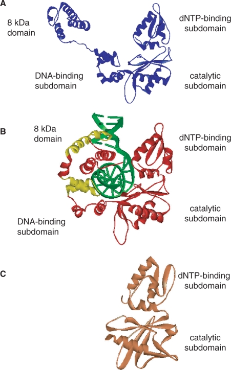
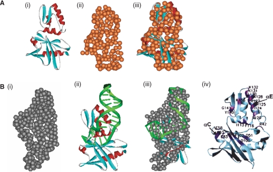
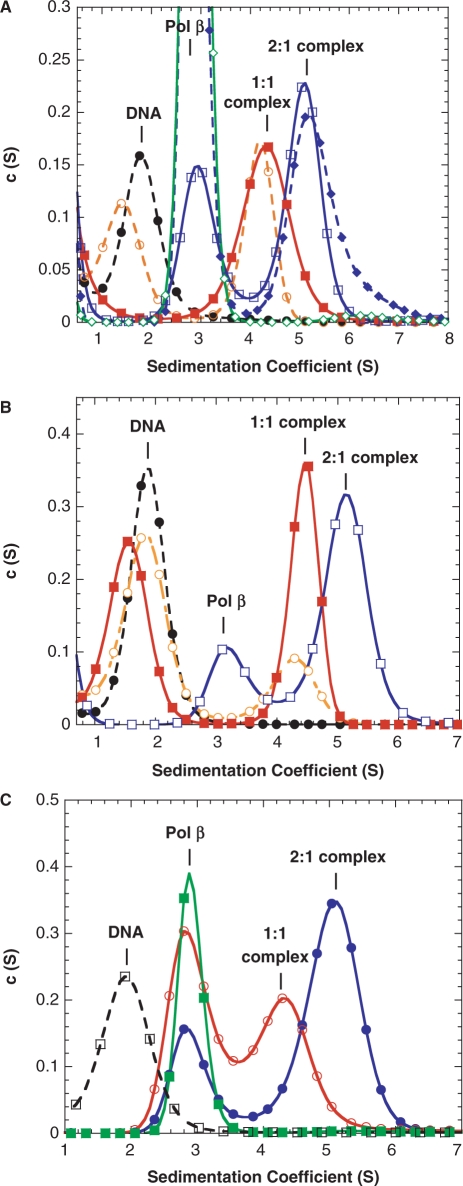
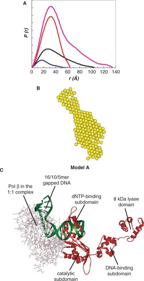
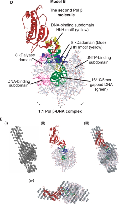
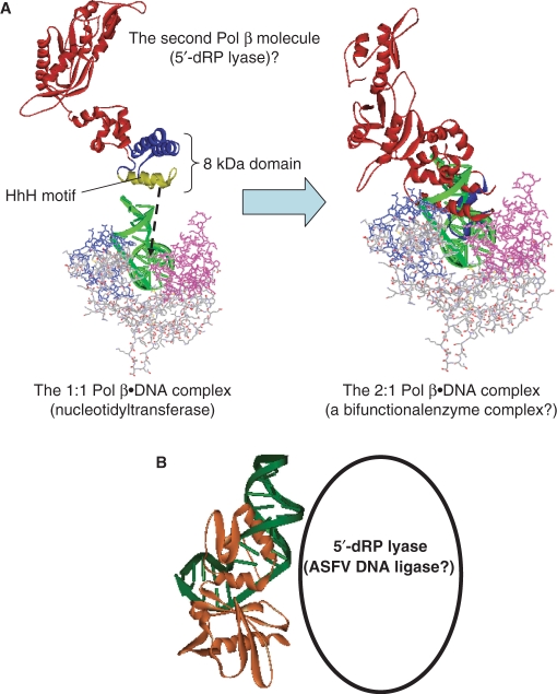
We report small-angle X-ray scattering (SAXS) and sedimentation velocity (SV) studies on the enzyme-DNA complexes of rat DNA polymerase beta (Pol beta) and African swine fever virus DNA polymerase X (ASFV Pol X) with one-nucleotide gapped DNA. The results indicated formation of a 2 : 1 Pol beta-DNA complex, whereas only 1 : 1 Pol X-DNA complex was observed. Three-dimensional structural models for the 2 : 1 Pol beta-DNA and 1 : 1 Pol X-DNA complexes were generated from the SAXS experimental data to correlate with the functions of the DNA polymerases. The former indicates interactions of the 8 kDa 5'-dRP lyase domain of the second Pol beta molecule with the active site of the 1 : 1 Pol beta-DNA complex, while the latter demonstrates how ASFV Pol X binds DNA in the absence of DNA-binding motif(s). As ASFV Pol X has no 5'-dRP lyase domain, it is reasonable not to form a 2 : 1 complex. Based on the enhanced activities of the 2 : 1 complex and the observation that the 8 kDa domain is not in an optimal configuration for the 5'-dRP lyase reaction in the crystal structures of the closed ternary enzyme-DNA-dNTP complexes, we propose that the asymmetric 2 : 1 Pol beta-DNA complex enhances the function of Pol beta.


Figures

Figure 1.
Figure 1.
The atomic resolution structures of Pol β and ASFV Pol X. (A) The crystal structure of free Pol β (1BPD); (B) the Pol β–DNA complex (1BPX). Protein and DNA moieties are shown in red and green, respectively, and the helix-hairpin-helix motifs are shown in yellow and (C) the NMR structure of free ASFV Pol X (1JAJ). The 8 kDa lyase domain, DNA-binding, catalytic and dNTP-binding subdomains are labeled. The 8 kDa lyase domain and DNA-binding subdomain in Pol β are absent in ASFV Pol X.
Figure 2.
Figure 2.
Structural studies of ASFV Pol X and ASFV Pol X–DNA complex. (A) (i) The NMR structure of the free ASFV Pol X (1JAJ), (ii) the GASBOR model derived from the SAXS data and (iii) superimpositions of the NMR structure and the GASBOR model. (B) The reconstructed model of the ASFV Pol X–DNA complex: (i) the DAMMIN model, (ii) the SASREF model, (iii) overlaid the DAMMIN and SASREF models and (iv) the predicted DNA-binding pocket (the residues involved are labeled) (9).
Figure 3.
Figure 3.
Comparisons of the experimental and calculated I(0) values from the DNA titration to Pol β. The calculated I(0) for the three-components model (free Pol β, free DNA and the 1:1 complex) (filled circle), the four-components model (free Pol β, free DNA, the 1:1 and 2:1 complexes) (filled triangle), no complex formation (open square), and the experimental I(0) for free Pol β(open diamond), free DNA (open triangle), and DNA titration to Pol β(filled square) are shown. The experimental and calculated data points are connected with bold and dash lines, respectively.
Figure 4.
Figure 4.
Sedimentation coefficient distribution plots for Pol β titration to DNA. (A) The experiments, monitored at 280 nm, were conducted at 1 μM DNA with 0 (filled circle, dashed lines), 1 (open circle, dashed lines), 3 (filled square, continuous lines), 9 (open square, dashed lines) and 27 (filled diamond, dashed lines) μM of Pol β. The c(S) data of 27 μM Pol β is shown in (open diamond, continuous lines). (B) The experiments, monitored at 260 nm, were conducted at 1 μM DNA with 0 (filled circle, dashed lines), 0.4 (open circle, dashed lines), 0.8 (filled square, continuous lines) and 7.2 (open square, continuous lines) μM Pol β. (C) The ionic strength effect studies were performed with the following samples: 1 μM DNA/9 μM Pol β with 0.1 (filled circle, continuous lines) and 0.4 M (open circle, continuous lines) KCl. The c(S) data of 9 μM Pol β in 0.1 M KCl (filled square, continuous lines) and 1 μM DNA in 0.4 M KCl (open square, dashed lines) are also plotted.
Figure 5.
Figure 5.
The reconstructed model of the 2:1 complex by SAXS. (A) The P(r) plots for free DNA (blue), free Pol β (black), the 1:1 complex (red) and 2:1 complex (magenta). (B) The DAMMIN model. (C) The SASREF model (model A) built with the ‘two-body’ approach (see text). (D) The SASREF model (model B) built with the ‘three-body’ approach (see text). The HhH motifs in the 1:1 complex and the second Pol β molecule are shown in pink and yellow, respectively. (E) (i) The DAMMIN model, (ii) the SASREF model (model B), (iii) and (iv) superimpositions of the DAMMIN and SASREF models are shown in two orthogonal views.
Figure 5.
Figure 5.
The reconstructed model of the 2:1 complex by SAXS. (A) The P(r) plots for free DNA (blue), free Pol β (black), the 1:1 complex (red) and 2:1 complex (magenta). (B) The DAMMIN model. (C) The SASREF model (model A) built with the ‘two-body’ approach (see text). (D) The SASREF model (model B) built with the ‘three-body’ approach (see text). The HhH motifs in the 1:1 complex and the second Pol β molecule are shown in pink and yellow, respectively. (E) (i) The DAMMIN model, (ii) the SASREF model (model B), (iii) and (iv) superimpositions of the DAMMIN and SASREF models are shown in two orthogonal views.
Figure 6.
Figure 6.
Proposed biological roles of the 2:1 Pol β–DNA complex and 1:1 ASFV Pol X–DNA complex in the BER pathway. (A) The HhH motifs in the 1:1 Pol β–DNA complex (lower left), the 8 kDa lyase domain of the second Pol β molecule (upper left), and the 2:1 Pol β–DNA complex (right) are highlighted in blue, and the fingers subdomain is labeled in pink. We propose that the first Pol β in the 2:1 complex functions as a nucleotidyl transferase, and the second Pol β molecule works as a 5′-deoxyribose phosphodiesterase interacting with the DNA damaged site. (B) Proposed interactions of a viral 5′-deoxyribose phosphodiesterase interacting with the ASFV Pol X–DNA complex in the viral BER pathway.

Similar articles

Cited by

References

    1. Sawaya MR, Pelletier H, Kumar A, Wilson SH, Kraut J. Crystal structure of rat DNA polymerase beta: evidence for a common polymerase mechanism. Science. 1994;264:1930–1935. - PubMed
    1. Steitz TA, Smerdon SJ, Jager J, Joyce CM. A unified polymerase mechanism for nonhomologous DNA and RNA polymerases. Science. 1994;266:2022–2025. - PubMed
    1. Tsoi PY, Yang M. Kinetic study of various binding modes between human DNA polymerase beta and different DNA substrates by surface-plasmon-resonance biosensor. Biochem. J. 2002;361:317–325. - PMC - PubMed
    1. Rajendran S, Jezewska MJ, Bujalowski W. Recognition of template-primer and gapped DNA substrates by the human DNA polymerase beta. J. Mol. Biol. 2001;308:477–500. - PubMed
    1. Bailey MF, Van der Schans EJ, Millar DP. Dimerization of the Klenow fragment of Escherichia coli DNA polymerase I is linked to its mode of DNA binding. Biochemistry. 2007;46:8085–8099. - PubMed

Publication types