. 2008 Feb;36(3):826-38.
doi: 10.1093/nar/gkm1107. Epub 2007 Dec 15.

Modulation of utrophin A mRNA stability in fast versus slow muscles via an AU-rich element and calcineurin signaling

Affiliations

Modulation of utrophin A mRNA stability in fast versus slow muscles via an AU-rich element and calcineurin signaling

Joe V Chakkalakal et al. Nucleic Acids Res. 2008 Feb.

Abstract

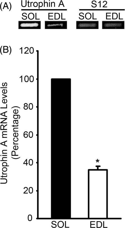
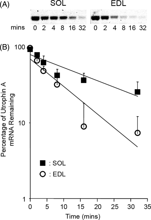
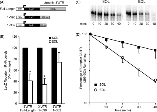
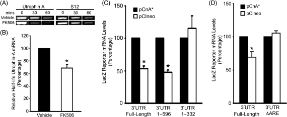
We examined the role of post-transcriptional mechanisms in controlling utrophin A mRNA expression in slow versus fast skeletal muscles. First, we determined that the half-life of utrophin A mRNA is significantly shorter in the presence of proteins isolated from fast muscles. Direct plasmid injection experiments using reporter constructs containing the full-length or truncated variants of the utrophin 3'UTR into slow soleus and fast extensor digitorum longus muscles revealed that a region of 265 nucleotides is sufficient to confer lower levels of reporter mRNA in fast muscles. Further analysis of this region uncovered a conserved AU-rich element (ARE) that suppresses expression of reporter mRNAs in cultured muscle cells. Moreover, stability of reporter mRNAs fused to the utrophin full-length 3'UTR was lower in the presence of fast muscle protein extracts. This destabilization effect seen in vivo was lost upon deletion of the conserved ARE. Finally, we observed that calcineurin signaling affects utrophin A mRNA stability through the conserved ARE. These results indicate that ARE-mediated mRNA decay is a key mechanism that regulates expression of utrophin A mRNA in slow muscle fibers. This is the first demonstration of ARE-mediated mRNA decay regulating the expression of a gene associated with the slow myogenic program.


Figures

Figure 1.
Figure 1.
Levels of utrophin A mRNA are higher in slow versus fast muscles. (A) Examples of ethidium bromide-stained agarose gels showing utrophin A and S12 PCR products from EDL and soleus (SOL) muscles. (B) Quantitation of utrophin A mRNA levels in EDL muscles expressed as a percentage of those found in SOL muscles and standardized to S12 transcripts. Note the significantly greater amount of utrophin A mRNA in SOL muscles. *Indicates a significant difference from EDL (P < 0.05; n = 4 independent experiments).
Figure 2.
Figure 2.
Utrophin A mRNA decays at a faster rate in the presence of fast muscle protein extracts. (A) In vitro stability assays were performed with protein extracts from EDL and soleus (SOL) muscles and RNA isolated from 3 day-old C2C12 myotubes. Examples of vistra green stained-agarose gels display utrophin A PCR products following 0, 2, 4, 8, 16 and 32 min incubation with protein extracts. Note the greater amount of utrophin A PCR products remaining upon incubation with the slow SOL muscle protein extracts versus with fast EDL muscle extracts. (B) Quantitation of the amounts of utrophin A mRNA at each time point, plotted in a semi-log scale as percentage of RNA seen at time 0. This analysis revealed that the half-lives seen in the presence of EDL versus soleus protein extracts were 3.55 min and 11.41 min, respectively (n = 4 independent experiments).
Figure 3.
Figure 3.
A region between nucleotides 332–596 in the utrophin 3′UTR decreases reporter mRNA expression in fast muscles. (A) Schematic representation of reporter constructs containing the full-length utrophin 3′UTR, and the first 596 and 332 inserted downstream of the LacZ reporter gene. (B) Mouse EDL and soleus (SOL) muscles were transduced with expression plasmids containing the various lengths of the 3′UTR. Note that LacZ reporter mRNA levels are ∼60% lower in EDL muscles when fused to either the utrophin full-length 3′UTR or the first 596 nucleotides. However, this difference is lost when LacZ mRNAs are fused to the first 332 nucleotides indicating the presence between nucleotides 332 and 596 of an instability element active in fast EDL muscles. *Indicates a significant difference from EDL (P < 0.05; n = 4–5 animals for each construct and in independent experiments). (C) Example of an in vitro stability assay using a radiolabeled RNA probe corresponding to nucleotide 299–603 of the utrophin 3′UTR incubated for various time periods with protein extracts from EDL and SOL muscles. (D) The intensity of the radioactive signals seen in these assays was quantified and plotted on a semi-log scale as a percentage of RNA seen at time 0 (n = 4 independent experiments). Note that the half-lives seen under these conditions were 17.36 min and 50.56 min when the radiolableld RNA probe was incubated with fast EDL versus slow SOL protein extracts, respectively.
Figure 4.
Figure 4.
The utrophin 3′UTR contains a conserved ARE that acts as a destabilizing element. (A) Sequence between nucleotides 332 and 596 of the utrophin 3′UTR. Grey characters denote three potential ARE. The underlined one is an ARE conserved between mouse and human which we subsequently deleted for further analysis. (B) Schematic representation of the utrophin full-length 3′UTR, the full-length version deleted of the conserved ARE (3′UTR ΔARE) and the SV40 3′UTR inserted downstream of the LacZ reporter construct. (C) Analysis of LacZ mRNA levels from 3 day-old C2C12 myotubes transfected with one of the three reporter constructs. Note the decrease in LacZ reporter mRNA expression when fused to the utrophin full-length 3′UTR relative to the SV40 3′UTR. Importantly, note also the loss of this effect upon deletion of the conserved ARE. *Indicates a significant difference relative to SV40 3′UTR, #indicates a significant difference relative to full-length 3′UTR [*(P < 0.05); #(P < 0.01) n = 3 independent experiments done in triplicate]. (D) Mouse EDL and soleus (SOL) muscles were injected with plasmids containing the utrophin 3′UTR ΔARE reporter constructs. Note that in contrast to injection with reporter constructs containing the utrophin full-length 3′UTR (Figure 3), expression levels of LacZ mRNA are not different between EDL and SOL muscles upon deletion of the conserved ARE (P > 0.05); n = 5 animals used in independent experiments.
Figure 5.
Figure 5.
The conserved ARE in the utrophin 3′UTR increases the rate of decay of reporter mRNAs in fast muscle. (A) Example of ethidium bromide-stained agarose gels showing LacZ PCR products obtained following an in vitro stability assay at 0, 30 and 60 min of incubation with EDL or soleus (SOL) protein extracts. Note the greater amount of LacZ mRNA fused to the utrophin full-length 3′UTR that remains after 30 and 60 min incubations with the slow SOL muscle protein extracts. Also, note the higher levels of LacZ mRNA remaining after 30 and 60 min incubations with EDL protein extracts upon deletion of the conserved ARE in the utrophin 3′UTR (compare full-length 3′UTR to 3′UTR ΔARE from EDL samples). (B) Quantitation of the relative half-life expressed as a percentage of the values obtained for LacZ mRNA fused to the utrophin full-length 3′UTR and incubated with SOL protein extracts. Note the significant decrease in the half-life for LacZ mRNA fused to the utrophin full-length 3′UTR upon incubation with EDL protein extracts relative to SOL. Also, note the significant increase in the half-life of LacZ mRNA upon deletion of the conserved ARE in the utrophin 3′UTR, relative to the utrophin full-length 3′UTR in samples incubated with EDL protein extracts. *Indicates a significant difference from SOL; #indicates a significant difference from other EDL samples (P < 0.05; n = 4 independent experiments).
Figure 6.
Figure 6.
Calcineurin signaling regulates the stability of utrophin A mRNA through its 3′UTR. In vitro stability assays were performed with soleus (SOL) protein extracts from mice treated with vehicle or the calcineurin inhibitor FK506, and RNA isolated from 3-day old C2C12 myotubes. (A) Example of ethidium bromide-stained agarose gels displaying utrophin A and S12 PCR products following 0, 30 and 60 min incubation with protein extracts. Note the greater amount of utrophin A mRNA remaining after 30 and 60 min incubations with SOL extracts from vehicle-treated mice as compared with SOL extracts from mice treated with FK506. (B) Quantitation of the relative half-life expressed as a percentage of values obtained for utrophin A incubated with SOL extracts from vehicle treated mice. Note the significant decrease in the half-life of utrophin A upon incubation with SOL extracts from FK506-treated mice. *Indicates a significant difference from vehicle (P < 0.05; n = 3 independent experiments). (C) C2C12 myoblasts were transfected with reporter constructs containing either the utrophin full-length 3′UTR, or the first 596 or 332 nucleotides (Figure 2A), together with an empty vector (pCIneo) or a construct containing a constitutively active variant of calcineurin (pCnA*). Note that LacZ reporter mRNA levels expressed from the utrophin full-length 3′UTR and the first 596 nucleotide constructs, are 50% higher in myotubes expressing CnA*. This difference is lost when LacZ mRNAs are fused to only the first 332 nucleotides indicating the presence of a calcineurin-responsive element between nucleotides 332 and 596. *Indicates a significant difference between pCIneo and pCnA*transduced muscles (P < 0.05; n = 4 experiments done in triplicate). (D) Tibilais anterior muscles were injected with plasmids containing the utrophin full-length 3′UTR or the utrophin 3′UTR ΔARE reporter constructs along with the pCIneo or pCnA*constructs. LacZ reporter mRNA levels from the full-length 3′UTR construct are significantly increased when muscles fibers have overexpressed pCnA*, whereas no increase is observed in muscle fibers injected with the 3′UTR ΔARE reporter construct. *Indicates a significant difference between pCIneo and pCnA*transduced muscles (P < 0.05; n = 3–4 mice).
Figure 7.
Figure 7.
Calcineurin signaling regulates utrophin 3′UTR reporter mRNA stability through the conserved ARE. In vitro stability assays were performed with soleus (SOL) extracts obtained from mice treated with vehicle or FK506, and RNA isolated from 3 day-old C2C12 myotubes transfected with reporter constructs containing the full-length 3′UTR or the ARE-deleted version (3′UTR ΔARE). (A) Example of ethidium bromide-stained agarose gels displaying LacZ PCR products following 0, 30 and 60 min incubation with protein extracts. Note the greater amount of LacZ mRNA fused to the utrophin full-length 3′UTR that remain after 30 and 60 min incubations with SOL extracts from vehicle-treated mice versus incubation with extracts from FK506-treated mice. Also, note the loss of this effect upon deletion of the conserved ARE. (B) Quantitation of the relative half-life expressed as a percentage of values obtained for LacZ mRNA fused to the utrophin full-length 3′UTR incubated with SOL protein extracts from vehicle-treated mice. Note the significant decrease in the half-life of LacZ mRNA with the full-length 3 UTR upon incubation with SOL extracts from FK506-treated mice relative to SOL extracts from vehicle-treated mice. Also, note the loss of this response to FK506 upon deletion of the ARE. *Indicates a significant difference from utrophin full-length 3′UTR (P < 0.05; n = 3 independent experiments).

Similar articles

Cited by

References

    1. Krag TO, Gyrd-Hansen M, Khurana TS. Harnessing the potential of dystrophin-related proteins for ameliorating Duchenne's muscular dystrophy. Acta Physiol. Scand. 2001;171:349–358. - PubMed
    1. Blake DJ, Weir A, Newey SE, Davies KE. Function and genetics of dystrophin and dystrophin-related proteins in muscle. Physiol. Rev. 2002;82:291–329. - PubMed
    1. Khurana TS, Hoffman EP, Kunkel LM. Identification of a chromosome 6-encoded dystrophin-related protein. J. Biol. Chem. 1990;265:16717–16720. - PubMed
    1. Nguyen TM, Ellis JM, Love DR, Davies KE, Gatter KC, Dickson G, Morris GE. Localization of the DMDL gene-encoded dystrophin-related protein using a panel of nineteen monoclonal antibodies: presence at neuromuscular junctions, in the sarcolemma of dystrophic skeletal muscle, in vascular and other smooth muscles, and in proliferating brain cell lines. J. Cell Biol. 1991;115:1695–1700. - PMC - PubMed
    1. Ohlendieck K, Ervasti JM, Matsumura K, Kahl SD, Leveille CJ, Campbell KP. Dystrophin-related protein is localized to neuromuscular junctions of adult skeletal muscle. Neuron. 1991;7:499–508. - PubMed

Publication types

MeSH terms