

Genome-wide tracking of unmethylated DNA Alu repeats in normal and cancer cells
- PMID: 18084025
- PMCID: PMC2241897
- DOI: 10.1093/nar/gkm1105
Genome-wide tracking of unmethylated DNA Alu repeats in normal and cancer cells
Abstract
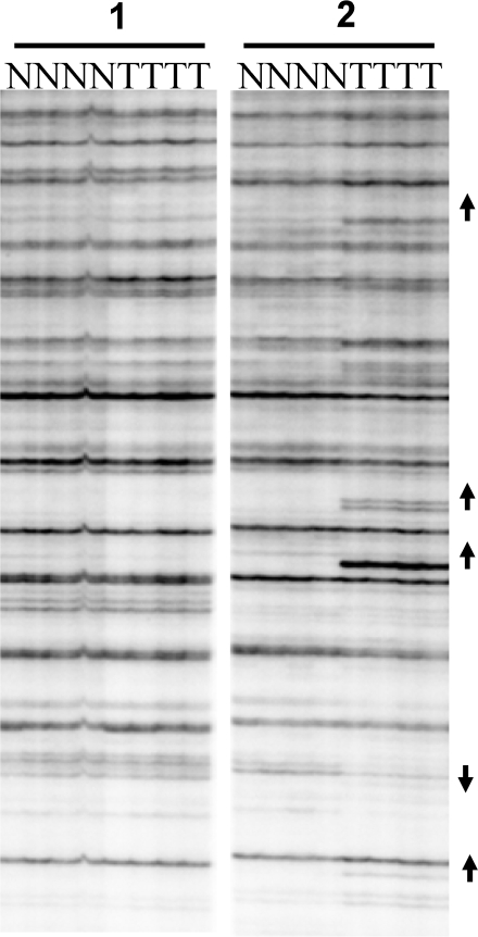
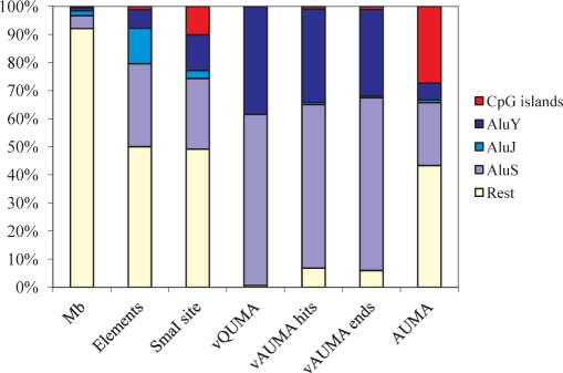
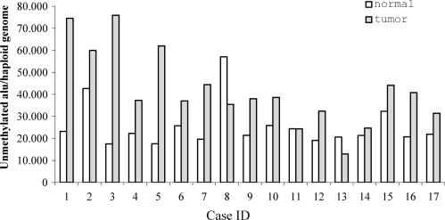
Methylation of the cytosine is the most frequent epigenetic modification of DNA in mammalian cells. In humans, most of the methylated cytosines are found in CpG-rich sequences within tandem and interspersed repeats that make up to 45% of the human genome, being Alu repeats the most common family. Demethylation of Alu elements occurs in aging and cancer processes and has been associated with gene reactivation and genomic instability. By targeting the unmethylated SmaI site within the Alu sequence as a surrogate marker, we have quantified and identified unmethylated Alu elements on the genomic scale. Normal colon epithelial cells contain in average 25 486 +/- 10 157 unmethylated Alu's per haploid genome, while in tumor cells this figure is 41 995 +/- 17 187 (P = 0.004). There is an inverse relationship in Alu families with respect to their age and methylation status: the youngest elements exhibit the highest prevalence of the SmaI site (AluY: 42%; AluS: 18%, AluJ: 5%) but the lower rates of unmethylation (AluY: 1.65%; AluS: 3.1%, AluJ: 12%). Data are consistent with a stronger silencing pressure on the youngest repetitive elements, which are closer to genes. Further insights into the functional implications of atypical unmethylation states in Alu elements will surely contribute to decipher genomic organization and gene regulation in complex organisms.
Figures

Similar articles
-
The epigenetic landscape of Alu repeats delineates the structural and functional genomic architecture of colon cancer cells.Genome Res. 2017 Jan;27(1):118-132. doi: 10.1101/gr.207522.116. Epub 2016 Dec 20. Genome Res. 2017. PMID: 27999094 Free PMC article.
-
High-throughput sequence-based epigenomic analysis of Alu repeats in human cerebellum.Nucleic Acids Res. 2009 Jul;37(13):4331-40. doi: 10.1093/nar/gkp393. Epub 2009 May 20. Nucleic Acids Res. 2009. PMID: 19458156 Free PMC article.
-
Alu element mutation spectra: molecular clocks and the effect of DNA methylation.J Mol Biol. 2004 Nov 26;344(3):675-82. doi: 10.1016/j.jmb.2004.09.058. J Mol Biol. 2004. PMID: 15533437
-
Alu elements and the human genome.Genetica. 2000;108(1):57-72. doi: 10.1023/a:1004099605261. Genetica. 2000. PMID: 11145422 Review.
-
[Alu repeats in the human genome].Mol Biol (Mosk). 2003 May-Jun;37(3):382-91. Mol Biol (Mosk). 2003. PMID: 12815945 Review. Russian.
Cited by
-
Hypomethylation coordinates antagonistically with hypermethylation in cancer development: a case study of leukemia.Hum Genomics. 2016 Jul 25;10 Suppl 2(Suppl 2):18. doi: 10.1186/s40246-016-0071-5. Hum Genomics. 2016. PMID: 27461342 Free PMC article.
-
Early detection of hepatocellular carcinoma via no end-repair enzymatic methylation sequencing of cell-free DNA and pre-trained neural network.Genome Med. 2023 Nov 8;15(1):93. doi: 10.1186/s13073-023-01238-8. Genome Med. 2023. PMID: 37936230 Free PMC article.
-
Epigenetic Regulation of microRNAs in Cancer: Shortening the Distance from Bench to Bedside.Int J Mol Sci. 2021 Jul 8;22(14):7350. doi: 10.3390/ijms22147350. Int J Mol Sci. 2021. PMID: 34298969 Free PMC article. Review.
-
Writing and rewriting the epigenetic code of cancer cells: from engineered proteins to small molecules.Mol Pharmacol. 2013 Mar;83(3):563-76. doi: 10.1124/mol.112.080697. Epub 2012 Nov 13. Mol Pharmacol. 2013. PMID: 23150486 Free PMC article. Review.
-
Chromosome 12p deletions in TEL-AML1 childhood acute lymphoblastic leukemia are associated with retrotransposon elements and occur postnatally.Cancer Res. 2008 Dec 1;68(23):9935-44. doi: 10.1158/0008-5472.CAN-08-2139. Cancer Res. 2008. PMID: 19047175 Free PMC article.
References
-
- Mattick JS. Challenging the dogma: the hidden layer of non-protein-coding RNAs in complex organisms. Bioessays. 2003;25:930–939. - PubMed
-
- Zuckerkandl E. Why so many noncoding nucleotides? The eukaryote genome as an epigenetic machine. Genetica. 2002;115:105–129. - PubMed
-
- Kreahling J, Graveley BR. The origins and implications of Aluternative splicing. Trends Genet. 2004;20:1–4. - PubMed
-
- Jasinska A, Krzyzosiak WJ. Repetitive sequences that shape the human transcriptome. FEBS Lett. 2004;567:136–141. - PubMed
-
- Jordan IK, Rogozin IB, Glazko GV, Koonin EV. Origin of a substantial fraction of human regulatory sequences from transposable elements. Trends Genet. 2003;19:68–72. - PubMed
Publication types
MeSH terms
LinkOut - more resources
Full Text Sources
Other Literature Sources
Medical
