

doi: 10.1107/S1744309107047781.
Epub 2007 Nov 21.
Hexammineruthenium(III) ion interactions with Z-DNA
Affiliations
- PMID: 18084080
- PMCID: PMC2344113
- DOI: 10.1107/S1744309107047781
Item in Clipboard
Hexammineruthenium(III) ion interactions with Z-DNA
Acta Crystallogr Sect F Struct Biol Cryst Commun.
.
Abstract
The hexamer duplex d(CGCGCA).d(TGCGCG) was crystallized with hexammineruthenium(III) ions in an orthorhombic space group; the crystals diffracted to 1.54 A resolution. Strong ion interactions with the adenine base induce a tautomeric shift from the amino to the imino form. Consequently, the A.T base pairing is disrupted. This structural study may be relevant to metal toxicity.
Figures
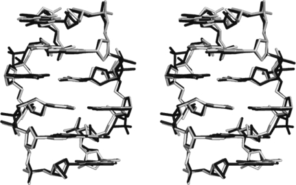
Least-squares superposition of the hexamer in the ruthenium complex (black) with the respective hexamer in the cobalt complex (grey).
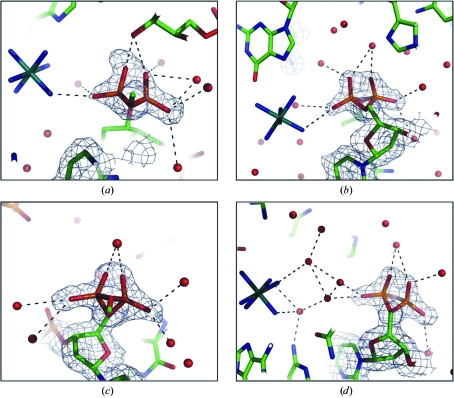
The conformation of the phosphate groups at residues C3 (a), C5 (b), C9 (c) and C11 (d); the respective 2F
obs − F
calc electron-density maps are contoured at the 1.0σ level. The dotted lines represent hydrogen bonds.
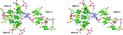
Stereoview of two base pairs from each of the three symmetry-related helices (A, x, y, z; B, −x + ½, −y, z + ½; C, x + ½, −y + ½, −z) interacting with the ion. The electron density shown for the ruthenium ion corresponds to the anomalous difference map.
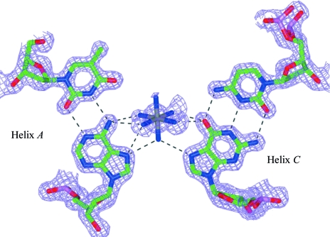
Ruthenium-ion interactions with helix A and its symmetry-related helix C. The electron-density map was calculated with (2F
obs − F
calc) as coefficients and contoured at the 1.0σ level.
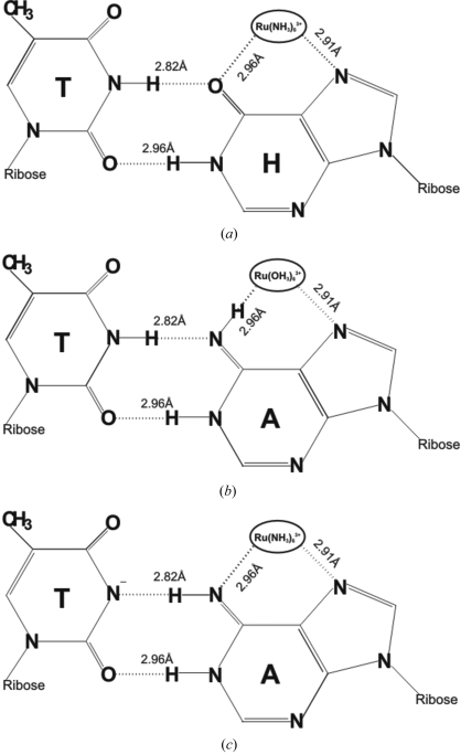
Schematic sketch of three possible explanations of ruthenium-ion interactions with the A6·T7 base pair. (a) Adenine is modified to hypoxanthine (H). (b) The amine groups of the ruthenium ion are exchanged with water. (c) The N3 imino proton of thymine is transferred to the environment.
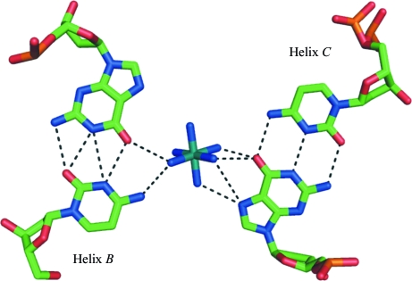
Geometry of the interaction of the ruthenium ion with helix B and helix C. The dotted lines represent hydrogen bonds.
Similar articles
-
Cobalt hexammine induced tautomeric shift in Z-DNA: the structure of d(CGCGCA)*d(TGCGCG) in two crystal forms.Nucleic Acids Res. 2004 Nov 8;32(19):5945-53. doi: 10.1093/nar/gkh919. Print 2004. Nucleic Acids Res. 2004. PMID: 15534365 Free PMC article.
-
Structure of d(TGCGCG).d(CGCGCA) in two crystal forms: effect of sequence and crystal packing in Z-DNA.Acta Crystallogr D Biol Crystallogr. 2005 Aug;61(Pt 8):1125-31. doi: 10.1107/S0907444905016781. Epub 2005 Jul 20. Acta Crystallogr D Biol Crystallogr. 2005. PMID: 16041078
-
Interaction between the Z-type DNA duplex and 1,3-propanediamine: crystal structure of d(CACGTG)2 at 1.2 A resolution.Biochemistry. 2006 Jan 31;45(4):1200-11. doi: 10.1021/bi051569l. Biochemistry. 2006. PMID: 16430216
-
Ultrahigh-resolution centrosymmetric crystal structure of Z-DNA reveals the massive presence of alternate conformations.Acta Crystallogr D Struct Biol. 2016 Nov 1;72(Pt 11):1203-1211. doi: 10.1107/S205979831601679X. Epub 2016 Oct 28. Acta Crystallogr D Struct Biol. 2016. PMID: 27841753
-
DNA architecture: from G to Z.Curr Opin Struct Biol. 2006 Jun;16(3):288-98. doi: 10.1016/j.sbi.2006.05.011. Epub 2006 May 22. Curr Opin Struct Biol. 2006. PMID: 16714104 Free PMC article. Review.
Cited by
-
Crystallization of Z-DNA in Complex with Chemical and Z-DNA Binding Z-Alpha Protein.Methods Mol Biol. 2023;2651:59-67. doi: 10.1007/978-1-0716-3084-6_4. Methods Mol Biol. 2023. PMID: 36892759
-
Predicting accurate ab initio DNA electron densities with equivariant neural networks.Biophys J. 2022 Oct 18;121(20):3883-3895. doi: 10.1016/j.bpj.2022.08.045. Epub 2022 Sep 3. Biophys J. 2022. PMID: 36057785 Free PMC article.
-
Phosphates in the Z-DNA dodecamer are flexible, but their P-SAD signal is sufficient for structure solution.Acta Crystallogr D Biol Crystallogr. 2014 Jul;70(Pt 7):1790-800. doi: 10.1107/S1399004714004684. Epub 2014 Jun 24. Acta Crystallogr D Biol Crystallogr. 2014. PMID: 25004957 Free PMC article.
-
High-resolution crystal structure of Z-DNA in complex with Cr(3+) cations.J Biol Inorg Chem. 2015 Apr;20(3):595-602. doi: 10.1007/s00775-015-1247-5. Epub 2015 Feb 17. J Biol Inorg Chem. 2015. PMID: 25687556 Free PMC article.
-
Interactions of Mn2+ with a non-self-complementary Z-type DNA duplex.Acta Crystallogr Sect F Struct Biol Cryst Commun. 2012 Dec 1;68(Pt 12):1420-6. doi: 10.1107/S1744309112041759. Epub 2012 Nov 14. Acta Crystallogr Sect F Struct Biol Cryst Commun. 2012. PMID: 23192018 Free PMC article.
References
-
- Alessio, E., Mestroni, G., Bergamo, A. & Sava, G. (2004). Curr. Top. Med. Chem.4, 1525–1535. - PubMed
-
- Brabec, V. (2002). Prog. Nucleic Acid Res. Mol. Biol.71, 1–68. - PubMed
-
- Brennan, R. G., Westhof, E. & Sundaralingam, M. (1986). J. Biomol. Struct. Dyn.3, 649–665. - PubMed
-
- Burda, J. V., Sponer, J. & Leszczynski, J. (2000). J. Biol. Inorg. Chem.5, 178–188. - PubMed
-
- Cate, J. H. & Doudna, J. A. (1996). Structure, 4, 1221–1229. - PubMed
Publication types
MeSH terms
Substances
Associated data
- Actions
- Actions
LinkOut - more resources
Full Text Sources
