

Production, purification and preliminary X-ray crystallographic studies of adeno-associated virus serotype 7
- PMID: 18084098
- PMCID: PMC2344100
- DOI: 10.1107/S1744309107060289
Production, purification and preliminary X-ray crystallographic studies of adeno-associated virus serotype 7
Abstract
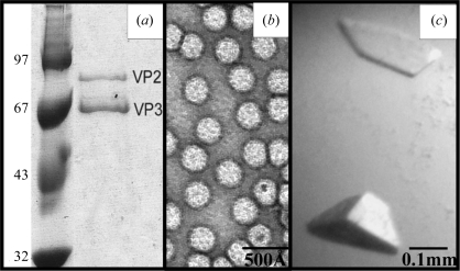
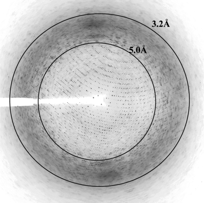
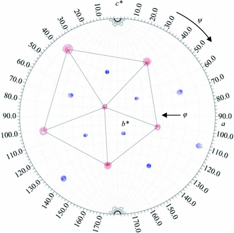
Crystals of baculovirus-expressed adeno-associated virus serotype 7 capsids diffract X-rays to approximately 3.0 A resolution. The crystals belong to the rhombohedral space group R3, with unit-cell parameters a = 252.4, c = 591.2 A in the hexagonal setting. The diffraction data were processed and reduced to an overall completeness of 79.0% and an R(merge) of 12.0%. There are three viral capsids in the unit cell. The icosahedral threefold axis is coincident with the crystallographic threefold axis, resulting in one third of a capsid (20 monomers) per crystallographic asymmetric unit. The orientation of the viral capsid has been determined by rotation-function searches and is positioned at (0, 0, 0) by packing considerations.
Figures
Similar articles
-
Production, purification and preliminary X-ray crystallographic studies of adeno-associated virus serotype 1.Acta Crystallogr Sect F Struct Biol Cryst Commun. 2006 Dec 1;62(Pt 12):1271-4. doi: 10.1107/S1744309106048184. Epub 2006 Nov 30. Acta Crystallogr Sect F Struct Biol Cryst Commun. 2006. PMID: 17142915 Free PMC article.
-
Production, purification, crystallization and preliminary X-ray analysis of adeno-associated virus serotype 8.Acta Crystallogr Sect F Struct Biol Cryst Commun. 2005 Jun 1;61(Pt 6):558-61. doi: 10.1107/S1744309105014132. Epub 2005 Jun 1. Acta Crystallogr Sect F Struct Biol Cryst Commun. 2005. PMID: 16511095 Free PMC article.
-
Production, purification and preliminary X-ray crystallographic studies of adeno-associated virus serotype 9.Acta Crystallogr Sect F Struct Biol Cryst Commun. 2009 Jul 1;65(Pt 7):715-8. doi: 10.1107/S1744309109021460. Epub 2009 Jun 27. Acta Crystallogr Sect F Struct Biol Cryst Commun. 2009. PMID: 19574648 Free PMC article.
-
Capsid-like arrays in crystals of chimpanzee adenovirus hexon.J Struct Biol. 2006 May;154(2):217-21. doi: 10.1016/j.jsb.2005.12.006. Epub 2006 Jan 18. J Struct Biol. 2006. PMID: 16458021 Review.
-
The role of the adeno-associated virus capsid in gene transfer.Methods Mol Biol. 2008;437:51-91. doi: 10.1007/978-1-59745-210-6_2. Methods Mol Biol. 2008. PMID: 18369962 Free PMC article. Review.
Cited by
-
AAV's anatomy: roadmap for optimizing vectors for translational success.Curr Gene Ther. 2010 Oct;10(5):319-340. doi: 10.2174/156652310793180706. Curr Gene Ther. 2010. PMID: 20712583 Free PMC article.
-
Twinned crystals of adeno-associated virus serotype 3b prove suitable for structural studies.Acta Crystallogr Sect F Struct Biol Cryst Commun. 2009 Feb 1;65(Pt 2):177-83. doi: 10.1107/S1744309109000372. Epub 2009 Jan 31. Acta Crystallogr Sect F Struct Biol Cryst Commun. 2009. PMID: 19194015 Free PMC article.
-
The structure of adeno-associated virus serotype 3B (AAV-3B): insights into receptor binding and immune evasion.Virology. 2010 Jul 20;403(1):26-36. doi: 10.1016/j.virol.2010.03.027. Epub 2010 May 4. Virology. 2010. PMID: 20444480 Free PMC article.
-
Perspective on Adeno-Associated Virus Capsid Modification for Duchenne Muscular Dystrophy Gene Therapy.Hum Gene Ther. 2015 Dec;26(12):786-800. doi: 10.1089/hum.2015.107. Epub 2015 Oct 15. Hum Gene Ther. 2015. PMID: 26414293 Free PMC article. Review.
-
Reengineered AAV vectors: old dog, new tricks.Discov Med. 2010 May;9(48):399-403. Discov Med. 2010. PMID: 20515607 Free PMC article.
References
-
- Brünger, A. T., Adams, P. D., Clore, G. M., DeLano, W. L., Gros, P., Grosse-Kunstleve, R. W., Jiang, J.-S., Kuszewski, J., Nilges, M., Pannu, N. S., Read, R. J., Rice, L. M., Simonson, T. & Warren, G. L. (1998). Acta Cryst. D54, 905–921. - PubMed
-
- Collaborative Computational Project, Number 4 (1994). Acta Cryst. D50, 760–763. - PubMed
-
- Emsley, P. & Cowtan, K. (2004). Acta Cryst. D60, 2126–2132. - PubMed
Publication types
MeSH terms
Substances
Grants and funding
LinkOut - more resources
Full Text Sources
Other Literature Sources
