. 2008 Jan;11(1):62-71.
doi: 10.1038/nn2027. Epub 2007 Dec 16.

A thermosensory pathway that controls body temperature

Affiliations

A thermosensory pathway that controls body temperature

Kazuhiro Nakamura et al. Nat Neurosci. 2008 Jan.

Abstract

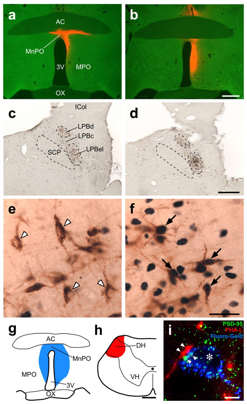
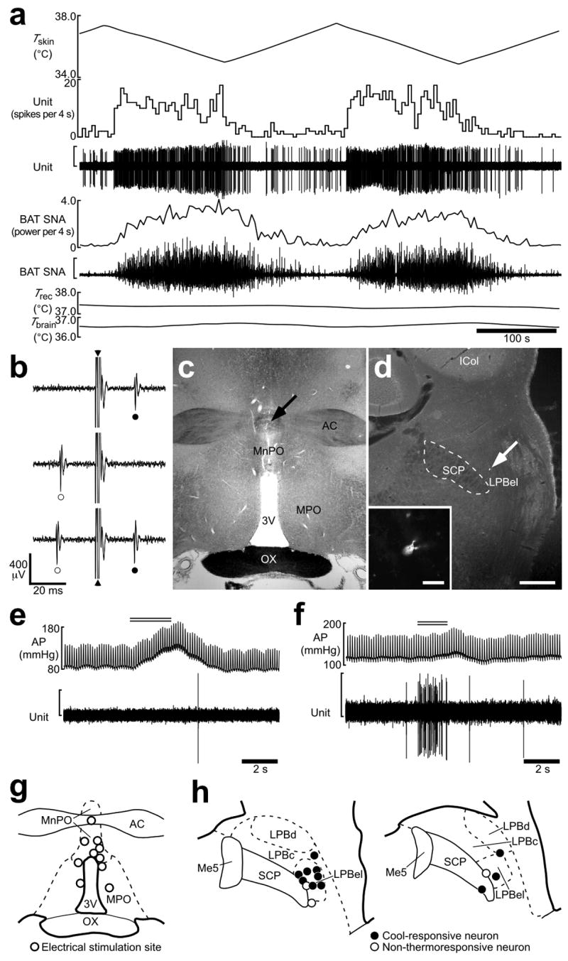
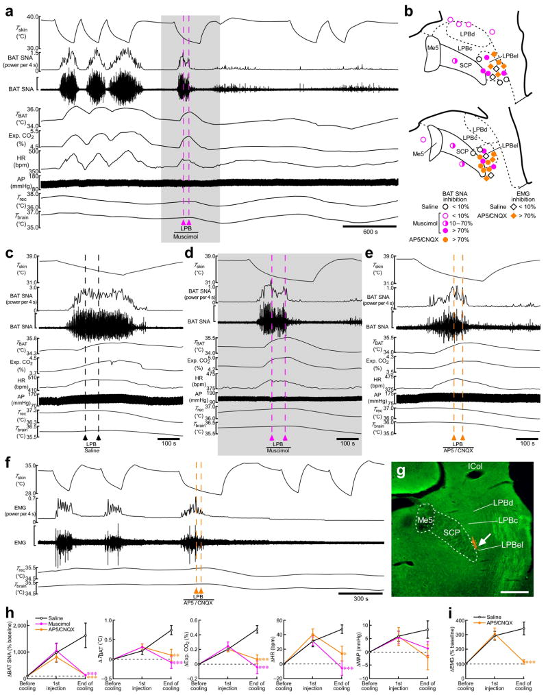
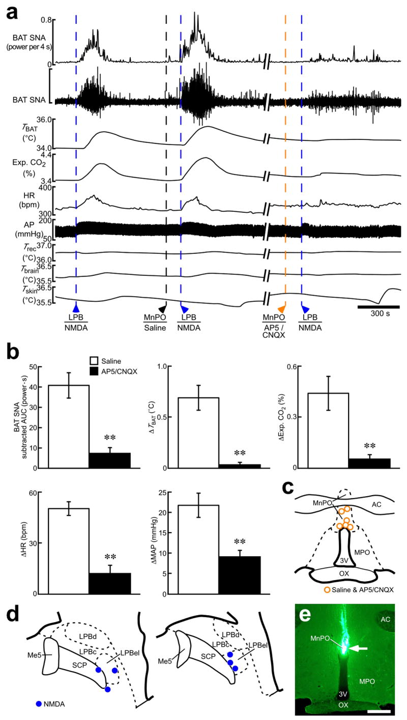
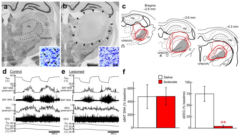
Defending body temperature against environmental thermal challenges is one of the most fundamental homeostatic functions that are governed by the nervous system. Here we describe a somatosensory pathway that essentially constitutes the afferent arm of the thermoregulatory reflex that is triggered by cutaneous sensation of environmental temperature changes. Using in vivo electrophysiological and anatomical approaches in the rat, we found that lateral parabrachial neurons are pivotal in this pathway by glutamatergically transmitting cutaneous thermosensory signals received from spinal somatosensory neurons directly to the thermoregulatory command center, the preoptic area. This feedforward pathway mediates not only sympathetic and shivering thermogenic responses but also metabolic and cardiac responses to skin cooling challenges. Notably, this 'thermoregulatory afferent' pathway exists in parallel with the spinothalamocortical somatosensory pathway that mediates temperature perception. These findings make an important contribution to our understanding of both the somatosensory system and thermal homeostasis -- two mechanisms that are fundamental to the nervous system and to our survival.


Figures

Figure 1
Figure 1
POA-projecting LPB neurons are activated in a cold environment and innervated by dorsal horn (DH) neurons. (af) Fos expression in LPB neurons retrogradely labeled with CTb injected into the POA in animals exposed to 24°C (a,c,e) and 4°C (b,d,f). a and b show injection sites of CTb (red). In cf, CTb (brown) and Fos (blue-black) immunoreactivities in the LPB of the animals shown in a and b were visualized. In e and f, arrowheads indicate Fos-negative, CTb-labeled neurons and arrows indicate Fos-positive, CTb-labeled neurons. 3V, third ventricle; AC, anterior commissure; ICol, inferior colliculus; MPO, medial preoptic area; OX, optic chiasm; SCP, superior cerebellar peduncle. Scale bars, 0.5 mm (ad), 30 μm (e,f). (gi) Dual tracing experiment using Fluoro-Gold (blue) injected into the POA (g) and PHA-L (red) injected into the spinal cervical DH (h). Confocal image from the LPBel (i) shows axon swellings, anterogradely labeled with PHA-L from the DH, closely associated (arrowheads) with PSD-95-positive (green) postsynaptic structures within LPB neurons retrogradely labeled with Fluoro-Gold from the POA. The signals for these three labelings are displayed using pseudo-colors. Asterisk indicates the cell nucleus of a Fluoro-Gold-labeled LPB neuron. VH, ventral horn. Scale bars, 0.5 mm (g,h), 5 μm (i).
Figure 2
Figure 2
Skin cooling-evoked response of single LPB neurons antidromically activated from the POA. (a) In vivo extracellular unit recording of the action potentials of an LPB neuron (unit) and changes in BAT SNA, rectal temperature (Trec) and brain temperature (Tbrain) in response to trunk skin cooling (Tskin). The vertical scale bars for the unit and BAT SNA traces represent 300 μV and 100 μV, respectively. Note that Trec and Tbrain do not change substantially during skin cooling and rewarming. (b) Collision test for the LPB neuron shown in a. Single pulse stimulation in the POA (triangle) evoked a constant-onset latency (20 ms) response in this neuron (filled circle, top trace). POA stimulation at 19 ms after a spontaneous action potential (open circle) failed to evoke a response of this neuron (middle trace). POA stimulation at 21 ms after a spontaneous action potential evoked a constant-onset latency response of this neuron (bottom trace). All traces are superpositions of 3 stimulation trials. (c) The site of electrical stimulation for the collision tests in b. The site is identified by a small scar at the site of electrical stimulation (arrow). (d) Juxtacellular labeling allows visualization of the LPB neuron (arrow) from a. Inset, a magnified picture of this neuron. Scale bars, 0.5 mm (c,d), 30 μm (inset in d). (e,f) Effect of tail pinch on firing activities of a cool-responsive neuron (e; the same neuron shown in a) and a non-thermoresponsive neuron (f). Double horizontal lines indicate the period of tail pinch. Note that tail pinch evoked a pressor response in both cases. The vertical scale bars for the unit traces in e and f represent 200 μV. (g) Sites of electrical stimulation in the POA. (h) Locations of LPB neurons that were juxtacellularly labeled after unit recording. Neurons antidromically activated with electrical stimulation in the POA are categorized in terms of their responsiveness to skin cooling. Me5, mesencephalic trigeminal nucleus.
Figure 3
Figure 3
Inhibition of neuronal activity or blockade of ionotropic glutamate receptors in the LPB reverses skin cooling-evoked thermogenic, metabolic and cardiac responses. (a) Skin cooling-evoked changes in BAT SNA, BAT temperature (TBAT), expired (Exp.) CO2, heart rate (HR), arterial pressure (AP), Trec, and Tbrain before and after bilateral nanoinjections of muscimol into the LPB (pink dashed lines). The gray area is expanded in d. The vertical scale bar for the BAT SNA trace represents 100 μV. (b) Composite drawing of sites of saline, muscimol (2 mM) or AP5/CNQX (5 mM each) nanoinjections (60 nl) in and around the LPB with their inhibitory effects on the skin cooling-evoked increase in BAT SNA or EMG. The right side of the symmetric bilateral injections is shown. (ce) Effect of bilateral nanoinjections of saline (c), muscimol (d) and AP5/CNQX (e) into the LPB on skin cooling-evoked changes in physiological variables. The vertical scale bar for the BAT SNA trace represents 200 μV (c), 100 μV (d) and 50 μV (e). (f) Skin cooling-evoked changes in EMG before and after bilateral nanoinjections of AP5/CNQX into the LPB (orange dashed lines). The vertical scale bar for the EMG trace represents 400 μV. g, Representative view of a nanoinjection site in the LPBel as identified with a cluster of fluorescent beads (arrow). Scale bar, 0.5 mm. (h,i) Group data (mean ± s.e.m.) showing the effect of saline (n = 5), muscimol (n = 7) or AP5/CNQX (n = 8) nanoinjections into the LPBel on skin cooling-evoked changes in BAT SNA, TBAT, Exp. CO2, HR and MAP (h) and the effect of saline (n = 4) and AP5/CNQX (n = 5) nanoinjections into the LPBel on skin cooling-evoked changes in EMG (i) (see cf). *P < 0.05; **P < 0.01; ***P < 0.001, compared with the saline-injected group (Bonferroni post hoc test following a one-way factorial ANOVA).
Figure 4
Figure 4
Stimulation of LPB neurons evokes thermogenic, metabolic and cardiovascular responses dependent on glutamatergic neurotransmission in the POA. (a) Effect of AP5/CNQX application (5 mM each, 100–200 nl) into the MnPO on thermogenic, metabolic and cardiovascular responses evoked by NMDA nanoinjection (0.2 mM, 36 nl) into the LPBel. The vertical scale bar for the BAT SNA trace represents 900 μV. (b) Group data (mean ± s.e.m.) showing the effect of pretreatment in the MnPO with saline or AP5/CNQX on increases in BAT SNA, TBAT, Exp. CO2, HR and MAP evoked by NMDA nanoinjection into the LPB. Each animal had an NMDA injection in the LPB following saline-pretreatment in the MnPO and then another NMDA injection in the LPB following AP5/CNQX-pretreatment in the MnPO and all the repeated injections were made at the same sites. **P < 0.01, n = 6 (two-tailed paired t-test). (c,d) Sites of injections of saline and AP5/CNQX into the MnPO (c) and of NMDA into the LPB (d). (e) Representative view of a nanoinjection site into the MnPO as identified with a cluster of fluorescent beads (arrow). Scale bar, 0.5 mm.
Figure 5
Figure 5
Skin cooling-evoked thermogenic response does not require a thalamic relay. (a,b) NeuN immunohistochemistry and cresyl violet staining (insets) in the thalamus of a control (a) and of a thalamic-lesioned (b) animal. Ibotenate injections eliminated neurons in an area including the VPM/VPL and posterior thalamic nuclear group (Po) (b, delineated by arrowheads) as compared with the saline-injected control (a). One side of the bilateral ibotenate or saline injection sites is shown. Large, neuron-like cells are found in the VPM/VPL of control animals (a, inset, arrows), but not of lesioned animals, which contained gliosis (b, inset). Scale bars, 1 mm (a,b), 30 μm (insets). (c) Thalamic area lesioned with ibotenate injections. Lesioned areas from all the animals are delineated with red lines and overlaid at three rostrocaudal levels. Gray area indicates the VPM/VPL. The right side of the bilateral symmetric lesions is shown. Hip, hippocampus; IC, internal capsule. (d,e) Skin cooling-evoked changes in BAT SNA and EEG in the animals from a and b. The vertical scale bars for the BAT SNA and EEG traces represent 25 μV and 200 μV (d) and 100 μV and 200 μV (e), respectively. (f) Group data (mean ± s.e.m.) showing the effect of the thalamic lesion on skin cooling-evoked changes in BAT SNA (saline, n = 5; ibotenate, n = 6) and EEG (saline, n = 3; ibotenate, n = 5). Skin cooling-evoked changes from the pre-cooling baseline to average value during the 1-min period immediately prior to the end of skin cooling (averaged from 2 cooling episodes in each animal) are shown. **P < 0.01 (two-tailed unpaired t-test).

Similar articles

Cited by

References

    1. Hammel HT. Regulation of internal body temperature. Annu Rev Physiol. 1968;30:641–710. - PubMed
    1. Hensel H. Thermoreception and temperature regulation. Monographs of the Physiological Society No. 38. Academic press; London: 1981. - PubMed
    1. Boulant JA, Gonzalez RR. The effect of skin temperature on the hypothalamic control of heat loss and heat production. Brain Res. 1977;120:367–372. - PubMed
    1. Nagashima K, Nakai S, Tanaka M, Kanosue K. Neuronal circuitries involved in thermoregulation. Auton Neurosci. 2000;85:18–25. - PubMed
    1. Nakamura K, Morrison SF. Central efferent pathways mediating skin cooling-evoked sympathetic thermogenesis in brown adipose tissue. Am J Physiol. 2007;292:R127–R136. - PMC - PubMed

Publication types

MeSH terms

Substances