. 2008 Jan;11(1):19-27.
doi: 10.1038/nn2026. Epub 2007 Dec 16.

MuSK controls where motor axons grow and form synapses

Affiliations

MuSK controls where motor axons grow and form synapses

Natalie Kim et al. Nat Neurosci. 2008 Jan.

Erratum in

  • Nat Neurosci. 2008 Feb;11(2):238

Abstract

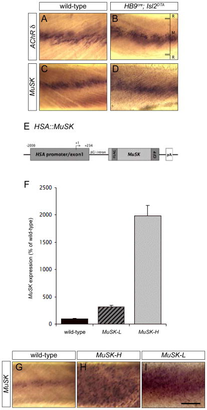
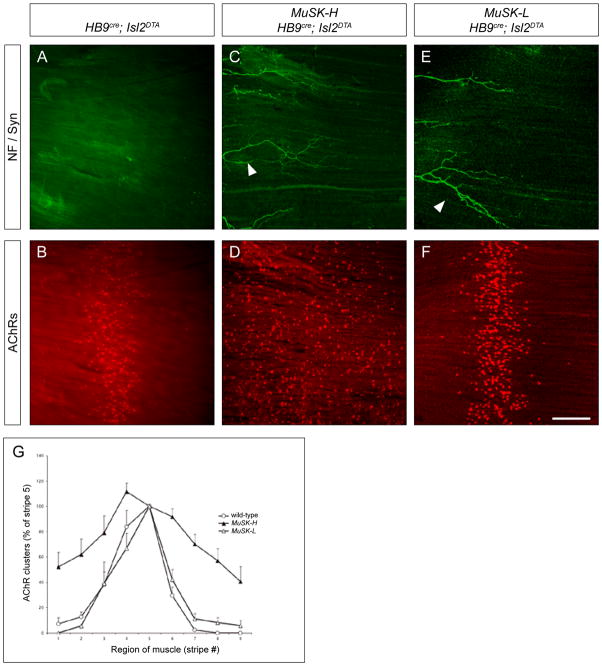
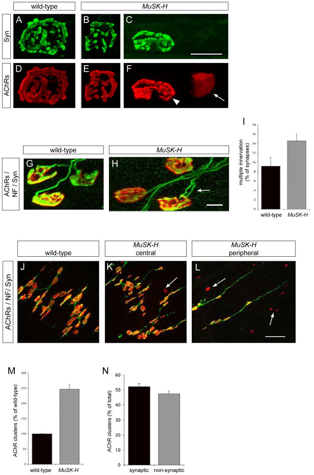
Motor axons approach muscles that are regionally prespecialized, as acetylcholine receptors are clustered in the central region of muscle before and independently of innervation. This muscle prepattern requires MuSK, a receptor tyrosine kinase that is essential for synapse formation. It is not known how muscle prepatterning is established, and whether motor axons recognize this prepattern. Here we show that expression of Musk is prepatterned in muscle and that early Musk expression in developing myotubes is sufficient to establish muscle prepatterning. We further show that ectopic Musk expression promotes ectopic synapse formation, indicating that muscle prepatterning normally has an instructive role in directing where synapses will form. In addition, ectopic Musk expression stimulates synapse formation in the absence of Agrin and rescues the lethality of Agrn mutant mice, demonstrating that the postsynaptic cell, and MuSK in particular, has a potent role in regulating the formation of synapses.


Conflict of interest statement

The authors do not have a conflict of interest related to this work.

Figures

Figure 1
Figure 1. Endogenous MuSK expression is patterned independent of innervation whereas HSA::MuSK is expressed uniformly in skeletal muscle
(A–D) Whole mounts of intercostal muscles from E18.5 wild-type (A, C) and HB9cre; Isl2DTA (B, D) embryos were processed for in situ-hybridization using probes for the AChR δ subunit and MuSK. MuSK mRNA (C, D) and AChR δ mRNA (A, B) are concentrated in the central region of muscle from wild-type and HB9cre; Isl2DTA mice, demonstrating that expression of endogenous MuSK is patterned in the absence of innervation. The positions of the ribs (R) and muscle (M) are indicated in B. (E) Schematic representation of the HSA::MuSK transgene. The human skeletal α-actin 5′ regulatory region was fused to a FLAG- and GFP-tagged MuSK cDNA. The FLAG tag was introduced into the extracellular domain of MuSK, and the GFP tag was added, in frame, to the MuSK carboxy terminus (Herbst et al. 2002). (F) The level of MuSK expression in two different HSA::MuSK transgenic lines was measured by real-time quantitative PCR. The low expressing line, MuSK-L, and the high expressing line, MuSK-H, express 3- and 20-fold more MuSK, respectively, than wild-type mice (MuSK-L: 311.29 ± 31.8 % of wild-type MuSK mRNA; MuSK-H: 1981.09 ± 197.2 % of wild-type MuSK; n = 4 mice for each genotype; the mean ± S.E.M.). (G–I) Whole mounts of intercostal muscles from P0 wild-type, MuSK-H and MuSK-L mice were processed for in situ hybridization to assess the pattern of MuSK transcription. The endogenous MuSK gene is transcribed selectively in the central, synaptic region of muscle (G), while the HSA regulatory region confers uniform MuSK expression (H, I). Scale bar = 200 μm.
Figure 2
Figure 2. A high level of ectopic MuSK induces ectopic AChR clusters and disrupts AChR prepatterning
(A–F) Whole mounts of diaphragm muscles from E18.5 mice that lack motor axons (A, B) and from mice that lack motor axons and carry a HSA::MuSK transgene (MuSK-H in C and D; MuSK-L in E and F), were stained with Alexa594-α-BGT (red) and antibodies to NF and Syn (green). Although motor axons are absent from muscle of HB9cre; Isl2DTA mice, the axons of sensory and/or autonomic neurons (arrowheads) are found at their normal location at the periphery of the muscle (A, C, E). AChR clusters are concentrated in the central region of muscle from HB9cre; Isl2DTA mice (B) but expressed throughout the muscle of mice that lack motor innervation and carry the MuSK-H transgene (D). Ectopic AChR clusters are not induced in muscle from mice that lack motor neurons and carry the MuSK-L transgene (F), showing that induction of ectopic AChR clusters is dependent upon the level of MuSK expression. (G) Quantitation of the distribution of AChR clusters (see Experimental Procedures). AChR clusters are concentrated in the central region of muscles from wild-type and MuSK-L mice, while AChR clusters are distributed throughout the muscle of MuSK-H mice (m ± S.E.M., n = 4 mice for each genotype). Scale bar for A–F = 200 μm.
Figure 3
Figure 3. A high level of ectopic MuSK promotes motor axon outgrowth
In wild-type mice (A, B), motor axons are fasciculated near the middle of the muscle (arrow), whereas the axons of sensory and/or autonomic neurons (arrowheads) are found at the periphery of the muscle. In wild-type mice, motor axons branch and terminate adjacent to the main intramuscular nerve (A), and AChRs are clustered selectively at synaptic sites (B). In mice carrying the MuSK-H transgene (C, D), the main intramuscular nerve is positioned correctly, but motor axons fail to stop and instead grow throughout the muscle (C). In contrast, in mice carrying the MuSK-L transgene (E, F), the pattern of motor axon outgrowth is indistinguishable from wild-type mice (E). (G) Quantitation of motor axon growth (see Experimental Procedures). Motor axons in wild-type and MuSK-L mice extend over 5–10% of the muscle, whereas motor axons in MuSK-H mice grow over 90% of the muscle (m ± S.E.M., n = 4 mice for each genotype). The expanded zone of axon growth found in P0 MuSK-H mice does not broaden further during postnatal development (Figure S1). Scale bar = 200 μm.
Figure 4
Figure 4. Ectopic MuSK induces ectopic synapses that arise from axon collateral branching
(A–C) Whole mounts of diaphragm muscles from wild-type (A) and MuSK-H (B) P0 mice were stained with Alexa594-α-BGT (red) and antibodies to NF and Syn (green). Anatomically matched areas from the left hemi-diaphragm were imaged from the medial to the costal edge of the muscle. In wild-type mice, synapses form only in the central region, adjacent to the main intramuscular nerve (A, D). In MuSK-H mice, synapses are distributed throughout the entire muscle (B, D). (C) Images of synapses in wild-type mice and in the central and peripheral regions of muscle from MuSK-H mice show that the general structure of ectopic synapses in MuSK-H appears normal. (D) The distribution of synapses was quantitated by dividing the muscle into 15 strips, and the number of synapses in each strip was determined. In wild-type mice, synapses are concentrated in a narrow central band that covers 5 to 10% of the muscle, while in MuSK-H muscle synapses are distributed throughout (~90%) the muscle (m ± S.E.M., n = 3 mice for each genotype). (E) Muscle from MuSK-H mice contains 28.76 ± 3.7% (mean ± S.E.M., n = 3 mice for each genotype) more synapses than muscle from wild-type mice. Synaptic size is lower in MuSK-H mice than in wild-type mice, whereas synaptic AChR density is similar in wild-type and MuSK-H mice (Figure S2). (F) Cross sections of diaphragm muscles from P0 MuSK-H and wild-type were stained with toluidine blue to count the number of muscle fibers. (G) The number of muscle fibers is similar in MuSK-H and wild-type mice (98.33± 4.6% of wild-type; n = 3 mice for each genotype; m ± S.E.M.). (H) Frozen sections from the cervical or lumbar region of the spinal cord, were stained with antibodies to Islet1/2 (green), and the number of motor neurons was determined (Experimental Procedures). (I) The number of lumbar spinal motor neurons is similar in MuSK-H and wild-type mice, as shown for L1-L5 (n = 3 mice for each genotype). The number of thoracic motor neurons is also similar in MuSK-H and wild-type mice (Figure S3). (J, K, L) We examined 495 synapses in diaphragm muscles from P11 MuSK-H (n = 3) and observed 4 instances of terminal sprouting (L). In contrast, we did not find a single instance of terminal sprouting in wild-type muscle (J). Scale bar = 200 μm for (A, B, F) and 10 μm for (C, J, K, L) and 100 μm for (H).
Figure 5
Figure 5. Synaptic AChR clusters mature, whereas ectopic, non-innervated AChR clusters remain simplified and are not extinguished in MuSK-H mice
(A–L) Whole mounts of diaphragm muscles from P60 (A–F), P11 (G, H) and P0 (J, K, L) wild-type and MuSK-H mice were stained with Alexa594-α-BGT (red) and antibodies to NF and Syn (green). Synaptic AChR clusters mature normally in MuSK-H mice, since they become perforated and highly branched (E), like in wild-type mice (D). Non-synaptic AChR clusters (arrow) do not mature but remain as ovoid plaques (arrowhead) (F). (G, H) Synapse elimination is delayed in MuSK-H mice. The number of multiply-innervated synapses (arrow) in the diaphragm muscle is modestly greater in MuSK-H than wild-type mice at P11 (H); by P30, all synapses are singly innervated in MuSK-H and wild-type mice (data not shown). (I) At P11, 14.6% ± 2% of synapses in MuSK-H and 9.17% ± 3.3% of synapses in wild-type mice are multiply-innervated (m ± S.E.M.). At least 140 synapses in 3 mice from each genotype were analyzed. (J, K, L) In P0 mice, non-innervated AChR clusters (arrows) are abundant in MuSK-H mice and more prevalent in the peripheral (L) than in the central region of the muscle (K). In contrast, all AChR clusters are innervated in muscle from P0 wild-type mice (J). (M) Muscle from MuSK-H mice express 2.5-fold more AChR clusters than wild-type mice (246.62 ± 15.0% of wild-type; n = 3 mice for each genotype; m ± S.E.M.). (N) In wild-type mice, each AChR cluster is innervated (data not shown), whereas in MuSK-H mice, 52.38 ± 1.8% of AChR clusters are innervated (n = 3 MuSK-H mice). Scale bar = 20 μm for (A–F) and 10 μm for (G, H) and 50 μm for (J, K, L).
Figure 6
Figure 6. The actin::MuSK-L transgene is sufficient to initiate but not maintain muscle prepatterning
Whole mounts of diaphragm muscles from E13.5 (A–C) or E18.5 (D–F) mice that lack (A, D) motor neurons (HB9cre; Isl2DTA), (B, E) motor neurons and endogenous MuSK (HB9cre; Isl2DTA; MuSK−/−) or (C, F) motor neurons and endogenous MuSK but carry the MuSK-L transgene (HB9cre; Isl2DTA; MuSK−/−; HSA::MuSK-L) were stained with Alexa594-α-BGT. AChRs are clustered in the central region of muscle from mice that lack motor axons (A, D) but not in muscle from mice that lack both motor axons and endogenous MuSK (B, E). (C) Expression of the MuSK-L transgene restores AChR clustering in the central region of muscle from E13.5 mice that lack motor neurons and endogenous MuSK, and the width of this zone is indistinguishable from control mice (G) (n = 3 HB9cre; Isl2DTA and 2 HB9cre; Isl2DTA; MuSK−/−; HSA::MuSK-L mice). By E18.5, however, this restricted pattern of AChR expression is lost, since AChR clusters are found throughout the muscle of mice that carry the MuSK-L transgene and lack motor neurons and endogenous MuSK. (H, I) A model for initiating and maintaining muscle prepatterning. (H) Muscles grow in length by the fusion of myoblasts at the ends of developing myotubes. The MuSK gene is activated early during myotube formation. Stochastic dimerization of MuSK protein in small myotubes leads to tyrosine phosphorylation and activation of MuSK. Activated MuSK stimulates two positive feedback loops, a post-translational feedback loop, which clusters MuSK protein, and a transcriptional feedback loop, which stimulates MuSK expression. Because MuSK activation occurs early during myotube formation and because muscles grow extensively from their ends, these feedback loops specialize the central region of the muscle and initiate and maintain muscle prepatterning. (I) The actin gene is likewise activated early during myotube formation, but the actin gene is uniformly expressed throughout muscle. Thus, in mice that lack endogenous MuSK and carry the actin::MuSK-L transgene, expression of MuSK protein is initiated early, leading to stimulation of the post-translational feedback loop, but MuSK RNA expression is not elevated in the central region of the muscle, since the actin promoter is not responsive to the transcriptional feedback loop. Thus, in these mice, muscle prepatterning is initiated but not maintained. Scale bar = 100 μm for (A–C) and 200 μm for (D–F).
Figure 7
Figure 7. Ectopic MuSK promotes synapse formation and stabilization in the absence of Agrin
In E18.5 mice lacking neural Agrin (agrinΔ zz) (A, B), or all forms of Agrin (agrin−/−) (G, H), AChR clusters are sparse, small (Figure S5) and infrequently contacted by motor axons (B and Figure S5). In contrast, in E18.5 mice that lack neural Agrin and carry either the MuSK-H (C, D) or MuSK-L transgene (E, F), larger AChR clusters are maintained in the muscle (Figure S5), and frequently contacted by motor axons that stop and differentiate as nerve terminals (D, F and Figure S5). Likewise, in mice that are null for Agrin and carry the MuSK-L transgene (I, J), AChR clusters are contacted by motor axons that differentiate as nerve terminals. Mice that lack neural Agrin die at birth, whereas mice that lack neural Agrin and carry either the MuSK-L or MuSK-H transgene survive postnatally for several weeks (K), demonstrating that the HSA::MuSK transgenes can restore functional synapses. Mice that lack all isoforms of Agrin and carry either the MuSK-L or MuSK-H transgene also survive postnatally (data not shown). The restoration of functional synapses by either MuSK transgene is also accompanied by a rescue in the level of AChR protein expression and in synapse-specific transcription (Figure S5). Scale bar = 200 μm.

References

    1. Arber S, Burden SJ, Harris AJ. Patterning of skeletal muscle. Curr Opin Neurobiol. 2002;12:100–103. - PubMed
    1. Bennett MR, Pettigrew AG. The formation of synapses in striated muscle during development. J Physiol. 1974;241:515–545. - PMC - PubMed
    1. Bennett MR, Pettigrew AG. The formation of neuromuscular synapses. Cold Spring Harb Symp Quant Biol. 1976;40:409–424. - PubMed
    1. Brennan KJ, Hardeman EC. Quantitative analysis of the human alpha-skeletal actin gene in transgenic mice. J Biol Chem. 1993;268:719–725. - PubMed
    1. Burden SJ. The formation of neuromuscular synapses. Genes Dev. 1998;12:133–148. - PubMed

Publication types

MeSH terms