

doi: 10.1038/nsmb1326.
Epub 2007 Dec 16.
Distinct binding modes specify the recognition of methylated histones H3K4 and H4K20 by JMJD2A-tudor
Affiliations
- PMID: 18084306
- PMCID: PMC2211384
- DOI: 10.1038/nsmb1326
Item in Clipboard
Distinct binding modes specify the recognition of methylated histones H3K4 and H4K20 by JMJD2A-tudor
Nat Struct Mol Biol.
2008 Jan.
Abstract
The lysine demethylase JMJD2A has the unique property of binding trimethylated peptides from two different histone sequences (H3K4me3 and H4K20me3) through its tudor domains. Here we show using X-ray crystallography and calorimetry that H3K4me3 and H4K20me3, which are recognized with similar affinities by JMJD2A, adopt radically different binding modes, to the extent that we were able to design single point mutations in JMJD2A that inhibited the recognition of H3K4me3 but not H4K20me3 and vice versa.
Figures
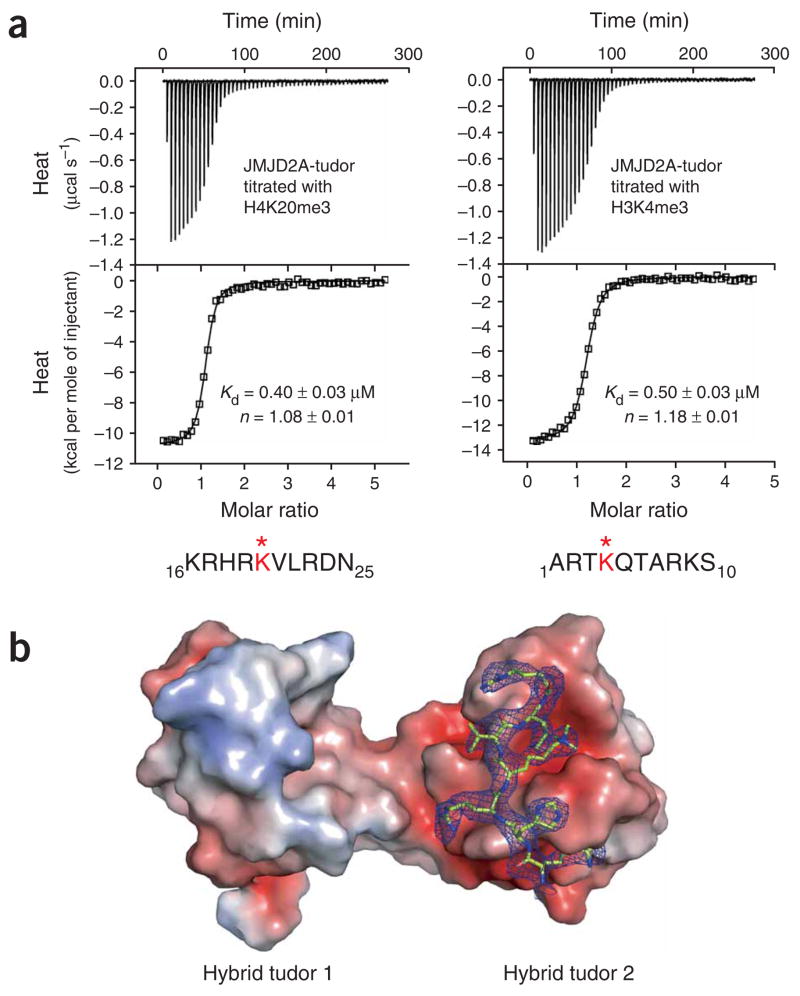
Interaction of JMJD2A hybrid tudor domains with trimethylated histones H3 and H4 peptides. (a) ITC of JMJD2A-tudor with H4K20me3 (left) and with H3K4me3 (right). The peptide amino acid sequences are indicated with the methylated (*) lysine in red. Raw titration data and integrated heat measurements are shown in the upper and lower plots, respectively. The Kd and stoichiometry numbers (n) obtained by fitting a standard one-interaction-site model are reported with the associated s.d. determined by nonlinear least-squares analysis. (b) Molecular surface and electrostatic potential representation of JMJD2A-tudor in complex with the H4K20me3 peptide. The electrostatic potential is shown in red for negatively charged and blue for positively charged surfaces. The peptide is in stick representation with the 2Fo– Fc electron density map displayed at the 1.0σ contour level.
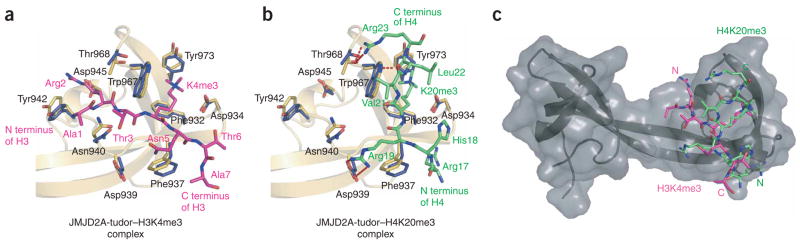
Two different binding modes of JMJD2A hybrid tudor domains with H3K4me3 and H4K20me3 peptides. (a, b) Close-up views of the JMJD2A-tudor interaction sites with H3K4me3 (a, PDB 2GFA6) and H4K20me3 (b). Amino acids of JMJD2A-tudor involved in binding H3K4me3 and H4K20me3 are shown. (c) Overall view of JMJD2A-tudor in complex with superimposed H3K4me3 (pink) and H4K20me3 (green), illustrating the opposite orientations of these peptides.
References
Publication types
MeSH terms
Substances
Associated data
- Actions
- Actions
Grants and funding
LinkOut - more resources
Full Text Sources
Molecular Biology Databases
