. 2008 Mar;153(5):936-46.
doi: 10.1038/sj.bjp.0707637. Epub 2007 Dec 17.

Endothelial alpha1-adrenoceptors regulate neo-angiogenesis

Affiliations

Endothelial alpha1-adrenoceptors regulate neo-angiogenesis

M Ciccarelli et al. Br J Pharmacol. 2008 Mar.

Abstract

Background and purpose: Intact endothelium plays a pivotal role in post-ischaemic angiogenesis. It is a phenomenon finely tuned by activation and inhibition of several endothelial receptors. The presence of alpha(1)-adrenoceptors on the endothelium suggests that these receptors may participate in regenerative phenomena by regulating the responses of endothelial cells involved in neo-angiogenesis.

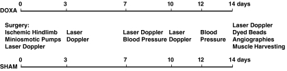
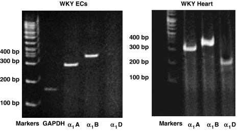
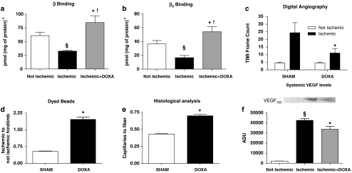
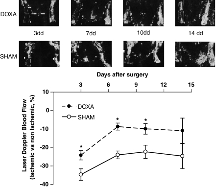
Experimental approach: We evaluated the expression of the subtypes of the alpha(1)-adrenoceptor in isolated endothelial cells harvested from Wistar-Kyoto (WKY) rats. We explored the possibility these alpha(1)-adrenoceptors may influence the pro-angiogenic phenotype of endothelial cells in vitro. In vivo, we used a model of hindlimb ischaemia in WKY rats, to assess the effects of alpha(1) adrenoceptor agonist or antagonist on angiogenesis in the ischaemic hindlimb by laser Doppler blood flow measurements, digital angiographies, hindlimb perfusion with dyed beads and histological evaluation.

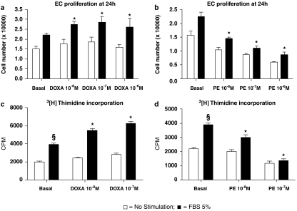
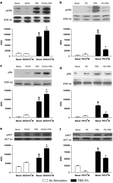
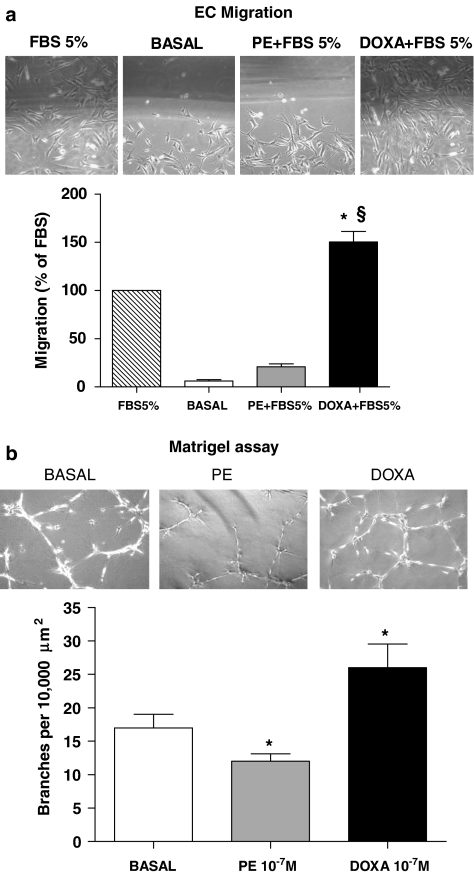
Key results: In vitro, pharmacological antagonism of alpha(1)-adrenoceptors in endothelial cells from WKY rats by doxazosin enhanced, while stimulation of these adrenoceptors with phenylephrine, inhibited endothelial cell proliferation and DNA synthesis, ERK and retinoblastoma protein (Rb) phosphorylation, cell migration and tubule formation. In vivo, we found increased alpha(1)-adrenoceptor density in the ischaemic hindlimb, compared to non-ischaemic hindlimb, suggesting an enhanced alpha(1)-adrenoceptor tone in the ischaemic tissue. Treatment with doxazosin (0.06 mg kg(-1) day(-1) for 14 days) did not alter systemic blood pressure but enhanced neo-angiogenesis in the ischaemic hindlimb, as measured by all our assays.

Conclusions: Our findings support the hypothesis that the alpha(1)-adrenoceptors in endothelial cells provide a negative regulation of angiogenesis.


Figures

Figure 1
Figure 1
Scheme of the in vivo experimental protocol.
Figure 2
Figure 2
Expression of α-adrenoceptor subtypes in Wistar-Kyoto (WKY) endothelial cells and WKY heart by reverse transcriptase-PCR. The figure shows that α1A- and α1B-adrenoceptors but not the α1D subtype were expressed in cultured rat aorta endothelial cells.
Figure 3
Figure 3
In vitro effects of doxazosin (DOXA; a, c) and phenylephrine (PE; b, d) on endothelial cell biology. All experiments depicted in this figure were performed from three to five times in duplicate. Role of increasing doses of doxazosin (a) and phenylephrine (b) on fetal bovine serum (FBS)-induced cell proliferation. Given alone, doxazosin did not affect endothelial cell proliferation. However, chronic incubation (24 h) with doxazosin enhanced endothelial cell proliferation in response to the mitogenic stimulus, FBS (5%; 24 h) (*P<0.05 vs basal+FBS) with a peak effect at a concentration of 10−7M doxazosin. In contrast, chronic incubation with phenylephrine reduced endothelial cell number and decreased proliferation (*P<0.05 vs basal+FBS). DNA synthesis assayed by [3H]thymidine incorporation. FBS (5%, 24 h) increased DNA synthesis (§P<0.03 vs basal); and this response was augmented by doxazosin (c) and reduced by phenylephrine (d) treatment (24 h) (*P<0.05 vs basal+FBS).
Figure 4
Figure 4
In vitro effects of doxazosin (DOXA) and phenylephrine (PE) on endothelial cell signal transduction. (a, b) Extracellular signal regulated kinase (ERK)/mitogen-activated protein kinase activation: western blot of activated (phosphorylated: pERK) ERK1/2 after stimulation with fetal bovine serum (FBS). Equal amounts of proteins were confirmed via blotting for total ERK. Representative blots are presented in the inset. Densitometric analysis (bar graph) shows that FBS stimulation caused ERK activation (§P<0.05 vs basal). Doxazosin alone did not increase ERK phosphorylation but significantly improved FBS-induced ERK activation (a). Phenylephrine 10−8M pre-incubation (24 h) did not change ERK activation but attenuated responses to FBS (b) (*P<0.05 vs basal+FBS; ANOVA; n=3–5 experiments, repeated in triplicate). (c, d) Progression in cell cycle evaluated by retinoblastoma phosphorylation (pRb). This protein regulates cell cycle progression through the restriction point within the G1 phase. After 12 h of stimulation with FBS. Rb was phosphorylated, as assessed by western blot (§P<0.05 vs basal). Densitometric analysis shows that doxazosin pre-incubation (10−7M, 24 h) enhanced Rb activation after FBS (c), whereas phenylephrine (10−8M, 24 h) reduced this response (d) (*P<0.05 vs basal+FBS). Equal amounts of proteins were verified by blotting for total ERK; n=3, repeated in duplicate. (e, f) Akt activation (pAkt) after FBS stimulation. FBS induced phosphorylation of Akt, doxazosin alone did not activate Akt, but hastened FBS activation (e). Conversely, phenylephrine (10−8M, 24 h) decreased FBS-induced activation of Akt (f) (*P<0.05 vs basal+FBS; ANOVA; n=3–5 experiments, repeated in triplicate). Equal amounts of proteins were verified by blotting for total Akt. ADU indicates arbitrary densitometry units, after correction for total protein content; representative blots are presented in the inset.
Figure 5
Figure 5
Cellular migration and vascular network formation. (a) Endothelial cell migration was measured 12 h after plating using a wounding assay. Migration of confluent endothelial cells was measured after the cell monolayer was partially wiped away. Photomicrographs show cells migrating into the wounded area. The area of the migrating cells was measured in several fields of view and is shown in the graph below. Data are presented as percent of migration with respect to fetal bovine serum (FBS) alone (*P<0.05 vs FBS 5%; §P<0.01 vs basal). (b) Endothelial cell network formation in vitro. Representative phase contrast photomicrographs of endothelial cells are shown plated on Matrigel in control conditions, in the presence of doxazosin 10−7M or phenylephrine 10−7M. Microscopy revealed numbers of network projections (branches) formed in each group after 12 h of incubation (*P<0.05 vs basal). It is interesting to note that phenylephrine modifies cell refraction, which is probably due to the favourable effects of phenylephrine on apoptosis. Data are presented as mean±s.e.
Figure 6
Figure 6
Increased neo-angiogenic responses by doxazosin (DOXA) treatment during chronic ischaemia in vivo. (a, b) β-Adrenoceptor density in rat hindlimbs. Total β-adrenoceptor (e) and β2-adrenoceptor (f) density were analysed in rat muscles from the ischaemic and non-ischaemic hindlimbs. We observed a reduction in both β-adrenoceptor and β2-adrenoceptor density within the ischaemic hindlimb (§P<0.03 vs not ischaemic). Rats receiving doxazosin in chronic infusion showed a significant upregulation of β- and β2-adrenoceptors (*P<0.02 vs ischaemic; !P<0.05 vs non-ischaemic). (c) TIMI frame count (TFC) of digital angiographies. This technique allows a better assessment of the deep vascular tree, which in the laser Doppler analysis is mostly affected by the cutaneous circulation. After 14 days of chronic ischaemia, digital angiographies showed a reduced number of TFCs in ischaemic hindlimbs treated with doxazosin, compared with sham rats (*P<0.05); the smaller TFC is indicative of improved blood perfusion. (d) Dyed beads dilution assay, where doxazosin treatment increased blood flow in ischaemic hindlimb, with respect to controls. Data shown are the dyed beads contained per milligram of hindlimb muscle tissue, expressed as the ratio between the ischaemic and non-ischaemic muscle (*P<0.05). (e) Histological analysis of capillaries in the rat tibialis anterior muscle. Compared with sham hindlimb, doxazosin increased capillary density, evaluated as number of capillaries corrected for number of muscle fibres, in the ischaemic tissue (*P<0.05). (f) Systemic levels of vascular endothelial growth factor (VEGF) in the non-ischaemic contralateral muscle. Fourteen days after femoral artery resection, we evaluated VEGF levels in the contralateral hindlimbs by western blots, as an indicator of systemic VEGF. Using muscle of rats that were not subjected to femoral artery resection as the non ischaemic reference (not ischaemic), we found that ischaemia caused an increase in systemic levels of VEFG (§P<0.01 vs non-ischaemic). Doxazosin treatment leads to a limitation of the ischaemic insult and consequently to a reduction of systemic levels of VEGF after 14 days (*P<0.05 vs ischaemic) (n=3, repeated in duplicate; a representative blot is presented in the inset; ADU: arbitrary densitometry units).
Figure 7
Figure 7
Laser Doppler analysis. Determination of laser Doppler blood flow on postoperative days 3, 7, 10, 14 shows a deficit in ischaemic hindlimb perfusion, compared with the contralateral hindlimb, that is significantly attenuated in doxazosin as compared with sham rats (*P<0.05, repeated measurements, ANOVA; laser Doppler blood flow data are expressed as percent of ischaemic to non-ischaemic limb).

Similar articles

Cited by

References

    1. Akhter SA, Luttrell LM, Rockman HA, Iaccarino G, Lefkowitz RJ, Koch WJ. Targeting the receptor–Gq interface to inhibit in vivo pressure overload myocardial hypertrophy. Science. 1998;280:574–577. - PubMed
    1. Alexandrov A, Keffel S, Goepel M, Michel MC. Stimulation of alpha1A-adrenoceptors in rat-1 cells inhibits extracellular signal-regulated kinase by activating p38 mitogen-activated protein kinase. Mol Pharmacol. 1998;54:755–760. - PubMed
    1. Augustin HG. Tubes, branches, and pillars: the many ways of forming a new vasculature. Circ Res. 2001;89:645–647. - PubMed
    1. Ben-Dov IZ, Ben-Arie L, Mekler J, Bursztyn M. How should patients treated with alpha-blockers be followed? Insights from an ambulatory blood pressure monitoring database. J Hypertens. 2006;24:861–865. - PubMed
    1. Benning CM, Kyprianou N. Quinazoline-derived alpha1-adrenoceptor antagonists induce prostate cancer cell apoptosis via an alpha1-adrenoceptor-independent action. Cancer Res. 2002;62:597–602. - PubMed

MeSH terms