. 2007 Dec;9(12):1066-77.
doi: 10.1593/neo.07667.

Inhibition of epidermal growth factor receptor and vascular endothelial growth factor receptor phosphorylation on tumor-associated endothelial cells leads to treatment of orthotopic human colon cancer in nude mice

Affiliations

Inhibition of epidermal growth factor receptor and vascular endothelial growth factor receptor phosphorylation on tumor-associated endothelial cells leads to treatment of orthotopic human colon cancer in nude mice

Takamitsu Sasaki et al. Neoplasia. 2007 Dec.

Abstract

The purpose of our study was to determine whether the dual inhibition of epidermal growth factor receptor (EGFR) and vascular endothelial growth factor receptor (VEGFR) signaling pathways in tumor-associated endothelial cells can inhibit the progressive growth of human colon carcinoma in the cecum of nude mice. SW620CE2 human colon cancer cells growing in culture and orthotopically in the cecum of nude mice expressed a high level of transforming growth factor alpha (TGF-alpha) and vascular endothelial growth factor (VEGF) but were negative for EGFR, human epidermal growth factor receptor 2 (HER2), and VEGFR. Double immunofluorescence staining revealed that tumor-associated endothelial cells expressed EGFR, VEGFR2, phosphorylated EGFR (pEGFR), and phosphorylated VEGFR (pVEGFR). Treatment of mice with either 7H-pyrrolo [2,3-d]-pyrimidine lead scaffold (AEE788; an inhibitor of EGFR and VEGFR tyrosine kinase) or CPT-11 as single agents significantly inhibited the growth of cecal tumors (P < .01); this decrease was even more pronounced with AEE788 combined with CPT-11 (P < .001). AEE788 alone or combined with CPT-11 also inhibited the expression of pEGFR and pVEGFR on tumor-associated endothelial cells, significantly decreased vascularization and tumor cell proliferation, and increased the level of apoptosis in both tumor-associated endothelial cells and tumor cells. These data demonstrate that targeting EGFR and VEGFR signaling on tumor-associated endothelial cells provides a viable approach for the treatment of colon cancer.

Keywords: AEE788; Endothelial cells; colon cancer; dual inhibition; tyrosine kinase receptors.


Figures
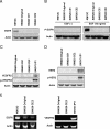
Figure 1
Figure 1
Expression of TGF-α, EGFR, VEGFR2, and HER2 by SW620CE2 cells growing in culture. Western blot and RT-PCR analyses reveal that the SW620CE2 cells do not express (A) EGFR, (B) HER2, or (C) VEGFR2 protein. KM12C human colon cancer cells served as a positive control for EGFR expression, and SKOV3ip1 human ovarian cancer cells served as a positive control for VEGFR2 and HER2 expression. The SW620CE2 cells produced high levels of (D) TGF-α but not (E) EGFR or VEGFR2 mRNA.
Figure 2
Figure 2
Western blot analysis for expression of phosphorylated Akt and phosphorylated ERK on murine endothelial cells. Murine endothelial cells established from ImmortoMouse were treated with AEE788 (1 µM) or vehicle solution for 1 hour and then stimulated for 15 minutes with serum-free DMEM, TGF-α (40 ng/ml), VEGF (40 ng/ml), or a combination of TGF-α and VEGF. Stimulation with TGF-α significantly increased the phosphorylation of Akt and ERK on endothelial cells. Stimulation with VEGF activated Akt but not ERK. The combination of TGF-α and VEGF produced additive activation of Akt and ERK. AEE788 treatment significantly inhibited the activation of Akt and ERK.
Figure 3
Figure 3
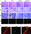
Immunohistochemical analysis of SW620CE2 cells growing in the cecum of nude mice. Double immunofluorescence staining was used for CD31/PECAM-1 and EGFR, pEGFR, VEGFR2, or pVEGFR2. (A) The SW620CE2 cells expressed TGF-α and VEGF but not EGFR or VEGFR. The KM12C cells expressed TGF-α and EGFR (green), and the SKOV3ip1 cells (growing in the peritoneal cavity) expressed VEGF and VEGFR2 (green). In all tumors, the endothelial cells expressed EGFR and VEGFR2 (yellow). (B) Colocalization for VEGFR1 and CD31 (endothelial cells) and F4/80 (macrophages) in SW620CE2 cecal tumors. (C) Expression of HER2 (green) in SKOV3ipl but not SW620CE2 tumors.
Figure 4
Figure 4
Gross pathology of SW620CE2 tumors in the cecum of nude mice.
Figure 5
Figure 5
Analysis for cell proliferation, apoptosis, and MVD. (A) Cecal tumors from different treatment groups underwent immunohistochemical analysis for the expression of CD31 (MVD), Ki-67 (cell proliferation), and apoptosis (TUNEL). Note that treatment with AEE788 alone decreased the number of CD31-positive and Ki-67 - positive cells and increased the number of TUNEL-positive cells; these changes were even more pronounced with the combination of AEE788 and CPT-11. (B) Double immunofluorescence staining was used for CD31/PECAM-1 (red) and TUNEL (green) in cecal tumors from mice treated with AEE788 and CPT-11 for only 2 weeks. The majority of apoptotic (TUNEL-positive) cells were endothelial cells.
Figure 6
Figure 6
Double immunofluorescence staining for expression of EGFR, pEGFR, VEGFR, or pVEGFR in tumor-associated endothelial cells. (A) Tissue sections were stained with anti -CD31/PECAM1 antibody (red) and with anti-EGFR, pEGFR, VEGFR, or pVEGFR (green) as described in the Materials and Methods section. Colocalization of CD31 and EGFR, pEGFR, VEGFR, or pVEGFR appears as yellow fluorescence. Expression of EGFR and VEGFR by tumor-associated endothelial cells was found in tumors from all treatment groups. Phosphorylation of EGFR and VEGFR on endothelial cells was inhibited by treatment with AEE788 or AEE788 plus CPT-11. Note that treatment with AEE788 alone decreased the number and diameter of blood vessels and that this effect was even more pronounced with combined AEE788 and CPT-11.

Similar articles

Cited by

References

    1. Lynch HT, de la Chapelle A. Hereditary colorectal cancer. N Engl J Med. 2003;348:919–932. - PubMed
    1. Bond JH. Colorectal cancer update: prevention, screening, treatment, and surveillance for high-risk groups. Med Clin North Am. 2000;84:1163–1182. - PubMed
    1. Fidler IJ. The organ microenvironment and cancer metastasis. Differentiation. 2002;70:498–505. (Review) - PubMed
    1. Fidler IJ. The pathogenesis of cancer metastasis: the “seed and soil” hypothesis revisited. Nat Rev Cancer. 2003;3:453–458. (Timeline) - PubMed
    1. Stoeltzing O, Liu W, Reinmuth N, Parikh A, Ahmad SA, Jung YD, Fan F, Ellis LM. Angiogenesis and antiangiogenic therapy of colon cancer liver metastasis. Ann Surg Oncol. 2003;10:722–733. - PubMed

Publication types

MeSH terms

LinkOut - more resources