. 2007 Dec;9(12):1078-90.
doi: 10.1593/neo.07727.

Statins stimulate in vitro membrane FasL expression and lymphocyte apoptosis through RhoA/ROCK pathway in murine melanoma cells

Affiliations

Statins stimulate in vitro membrane FasL expression and lymphocyte apoptosis through RhoA/ROCK pathway in murine melanoma cells

Guillaume Sarrabayrouse et al. Neoplasia. 2007 Dec.

Abstract

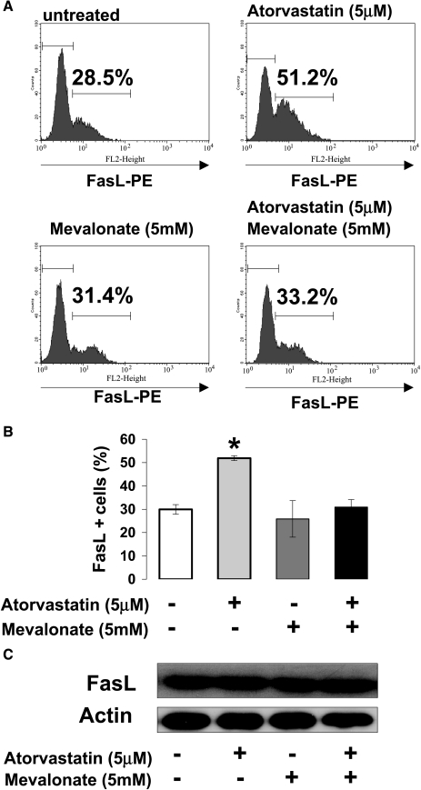
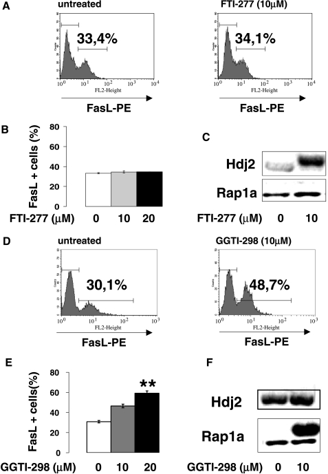
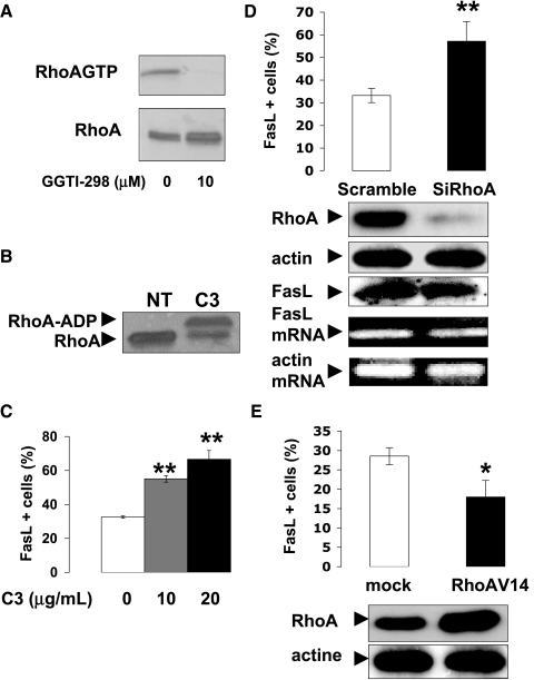
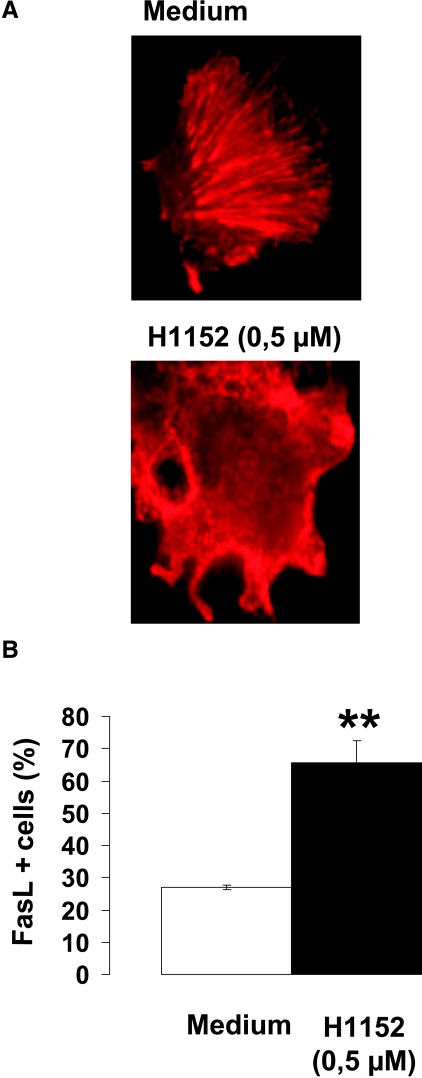
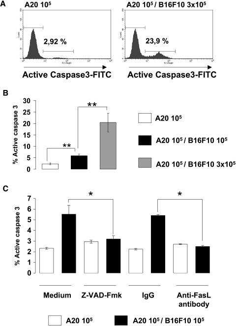
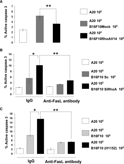
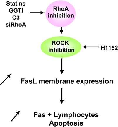
The capacity of FasL molecules expressed on melanoma cells to induce lymphocyte apoptosis contributes to either antitumor immune response or escape depending on their expression level. Little is known, however, about the mechanisms regulating FasL protein expression. Using the murine B16F10 melanoma model weakly positive for FasL, we demonstrated that in vitro treatment with statins, inhibitors of 3-hydroxy-3-methylgutaryl CoA reductase, enhances membrane FasL expression. C3 exotoxin and the geranylgeranyl transferase I inhibitor GGTI-298, but not the farnesyl transferase inhibitor FTI-277, mimic this effect. The capacity of GGTI-298 and C3 exotoxin to inhibit RhoA activity prompted us to investigate the implication of RhoA in FasL expression. Inhibition of RhoA expression by small interfering RNA (siRNA) increased membrane FasL expression, whereas overexpression of constitutively active RhoA following transfection of RhoAV14 plasmid decreased it. Moreover, the inhibition of a RhoA downstream effector p160ROCK also induced this FasL overexpression. We conclude that the RhoA/ROCK pathway negatively regulates membrane FasL expression in these melanoma cells. Furthermore, we have shown that B16F10 cells, through the RhoA/ROCK pathway, promote in vitro apoptosis of Fas-sensitive A20 lymphoma cells. Our results suggest that RhoA/ROCK inhibition could be an interesting target to control FasL expression and lymphocyte apoptosis induced by melanoma cells.

Keywords: FasL; Melanoma; RhoA; apoptosis; statins.


Figures

Figure 1
Figure 1
Statins upregulate FasL expression on B16F10 cell membrane. B16F10 melanoma cells were incubated with atorvastatin (5 µM) mevalonate (5 mM), or both for 48 hours. Cells were harvested and FasL was immunodetected by flow cytometry using a phycoerythrin-conjugated murine FasL monoclonal antibody. The percentage of FasL-positive cells (FasL+ cells (%)) is indicated (A). This percentage of FasL-positive cells was also analyzed in three independent experiments (B). A statistically significant increase (P < .05) is illustrated by an asterisk (*). Similarly treated B16F10 cells were lysed and immunoblotted with murine anti-FasL antibody for total FasL protein detection. Data from one representative of three independent experiments are shown (C).
Figure 2
Figure 2
Inhibition of protein geranylgeranylation by GGTI-298 increases FasL expression on B16F10 cell membrane. B16F10 cells were incubated with FTI-277 (10 and 20 µM) or GGTI-298 (10 and 20 µM) for 48 hours. Cells were harvested and FasL was immunodetected by flow cytometry. The percentage of FasL-positive cells is indicated (A and D). Histograms illustrating this percentage of FasL+ cells analyzed by flow cytometry from three independent experiments, using increasing doses of FTI-277 (0, 10, and 20 µM) (B) or GGTI-298 (0, 10, and 20 µM) (E). A statistically significant increase (P < .01) is illustrated by double asterisks (**). Similarly treated B16F10 cells were also analyzed and immunoblotted with Rap1a or Hdj2 antibody to confirm farnesyl transferase inhibition (C) and geranylgeranyl transferase inhibition (F) obtained by FTI-277 (10 µM) or GGTI-298 (10 µM) treatment, respectively.
Figure 3
Figure 3
RhoA negatively regulates membrane FasL expression on B16F10 melanoma cells. (A) B16F10 cells were incubated with GGTI-298 (10 µM) for 48 hours and RhoA activity was analyzed by TRBD assay as described in the Materials and Methods section. (B) B16F10 cells were incubated with C3 exoenzyme (10 µg/ml) for 48 hours. Untreated and treated cells were lysed and immunoblotted with anti-RhoA antibody to test RhoA-ADP ribosylation as described in the Materials and Methods section. (C) B16F10 cells were treated with C3 exoenzyme (10 and 20 µg/ml). Cells were harvested and FasL was immunodetected by flow cytometry. The percentage of FasL-positive cells (FasL+ cells (%)) was analyzed; data shown are mean values of three independent and reproducible experiments. A statistically significant increase (P < .01) is illustrated by double asterisks (**). (D) B16F10 cells were transfected either with scramble siRNA or with RhoA-specific siRNA (SiRhoA) and analyzed after 72 hours to evaluate by cytofluorometry the percentage of FasL-positive cells. The scramble siRNA contains a disorganized sequence of the same nucleotides and is therefore unable to cut the target RNA sequence. Again, data shown are mean values of three independent and reproducible experiments. A statistically significant increase (P < .01) is illustrated by double asterisks (**). Similarly transfected cells were also lysed and immunoblotted with either anti-RhoA antibody or anti-FasL antibody and compared to actin expression to check the efficiency of the RhoA-specific siRNA and the total FasL protein expression. RNA of these transfected cells were also used to compare their FasL mRNA levels by RT-PCR, as described in the Materials and Methods section. (E) B16F10 cells were also transfected with a pSG5 empty vector (Mock) or with a pSG5-RhoAV14 vector encoding for the active form of RhoA (RhoAV14). After 72 hours, membrane-expressed FasL was immunodetected by flow cytometry. The percentages of FasL-positive cells are shown as mean values of three independent and reproducible experiments. A statistically significant decrease (P < .05) is illustrated by an asterisk (*). These cells were also lysed and immunoblotted with anti-RhoA antibody to test the efficiency of the transfected pSG5-RhoAV14 vector to increase RhoA expression in B16F10 cells.
Figure 4
Figure 4
Inhibition of p160ROCK increases FasL expression on B16F10 cell membrane. (A) B16F10 cells were seeded onto glass coverslips in six-well plates to obtain 60% confluence on day 2. On day 1, cells were treated with 0.5 µM H1152 for 24 hours. After treatment, actin fibers were visualized by tetramethylrhodamine isothiocyanate-labeled phaloidin. Cells were viewed under a Zeiss Axiophot microscope (x 630), and pictures taken with a Princeton Camera. (B) B16F10 cells were incubated with 0.5 µM H1152 for 24 hours. Cells were harvested and FasL was immunodetected by flow cytometry. Histograms illustrating the percentage of FasL+ cells analyzed by flow cytometry from three independent experiments. A statistically significant increase (P < .01) is illustrated by double asterisks (**).
Figure 5
Figure 5
Apoptosis induced by Fas/FasL pathway of A20 lymphoma cocultivated with B16F10 melanoma cells. (A) A total of 1 x 10⁵ A20 cells were cultivated alone or with 3 x 10⁵ of B16F10 melanoma cells for 24 hours and the expression of the active form of caspase-3 was evaluated by cytofluorometry with a specific antibody in permeabilized A20 cells. Data shown are from one representative of three independent and reproducible experiments. (B) A total of 1 x 10⁵ A20 cells were cultivated alone or with increasing numbers of B16F10 melanoma cells (1 x 10⁵; 3 x 10⁵) for 24 hours and expression of the active form caspase-3 was evaluated by cytofluorometry. Data shown are mean values of three independent experiments. A statistically significant increase (P < .01) is illustrated by double asterisks (**). (C) A total of 1 x 10⁵ A20 cells were also cultivated for 24 hours alone or with 1 x 10⁵ B16F10 cells, in the absence (medium) or the presence of 100 µM caspase inhibitor Z-VAD-Fmk (Z-VAD-Fmk). Medium corresponds to the normal culture medium containing 1% DMSO to be in the same conditions as those required by the Z-VAD-Fmk inhibitor. Coculture experiments were also carried out with blocking anti-FasL antibody (c178) at 50 µg/ml (anti-FasL antibody) or matched isotype control (IgG). IgG corresponds to the normal culture medium containing 50 µg/ml of a rabbit IgG used as control for the anti-FasL antibody. Data shown are mean values of three independent experiments. Statistically significant decreases (P < .05) induced by the inhibitor or the blocking antibody are illustrated by an asterisk (*).
Figure 6
Figure 6
RhoA negatively regulates FasL-induced apoptosis of A20 cells cocultivated with B16F10 melanoma cells. (A) A20 lymphoma cells (1 x 10⁵) were cultivated for 24 hours, either alone or with 1 x 10⁵ B16F10 cells, transfected with either pSG5 empty vector (B16F10Mock) or pSG5-RhoAV14 vector (B16F10RhoAV14). The percentage of A20 cells expressing the active form of caspase-3 (% active caspase-3) was analyzed by flow cytometry. (B) A20 lymphoma cells (1 x 10⁵) were also cocultivated for 24 hours either alone or with 1 x 10⁵ B16F10 cells transfected with a control siRNA (B16F10Sc) or a RhoA-specific siRNA (B16F10SiRhoA). (C) A20 cells (1 x 10⁵) were also cocultivated for 24 hours either alone or with 1 x 10⁵ B16F10 cells treated or not by ROCK inhibitor (0.5 µM H1152). These cultures (B) and (C) were made both in a control medium containing an IgG isotype control (IgG) and in a medium containing a blocking anti-FasL antibody at 50 µ g/ml (antibody anti-FasL). The percentage of A20 cells expressing the active form of caspase-3 was analyzed by flow cytometry. Data shown are from one experiment representative of three. Statistically significant increases or decreases of A20 cells expressing the active form of caspase-3 are illustrated by an asterisk (*) (P < .05) or double asterisks (**) (P < .01).
Figure 7
Figure 7
Schematic representation of the effects of RhoA/ROCK signaling pathway inhibition on membrane FasL expression and lymphocytes apoptosis. In vitro treatments of B16F10 melanoma cells with inhibitors of the RhoA/ROCK pathway increase the membrane FasL expression on these treated tumor cells. This (over)expression promoted the apoptosis of Fas-sensitive lymphocytes.

Similar articles

Cited by

References

    1. Suda T, Takahashi T, Golstein P, Nagata S. Molecular cloning and expression of the Fas ligand, a novel member of the tumor necrosis factor family. Cell. 1993;75:1169–1178. - PubMed
    1. Trauth BC, Klas C, Peters AM, Matzku S, Moller P, Falk W, Debatin KM, Krammer PH. Monoclonal antibody-mediated tumor regression by induction of apoptosis. Science. 1989;245:301–305. - PubMed
    1. Los M, Wesselborg S, Schulze-Osthoff K. The role of caspases in development, immunity, and apoptotic signal transduction: lessons from knockout mice. Immunity. 1999;10:629–639. - PubMed
    1. Itoh N, Yonehara S, Ishii A, Yonehara M, Mizushima S, Sameshima M, Hase A, Seto Y, Nagata S. The polypeptide encoded by the cDNA for human cell surface antigen Fas can mediate apoptosis. Cell. 1991;66:233–243. - PubMed
    1. Yonehara S, Ishii A, Yonehara M. A cell-killing monoclonal antibody (anti-Fas) to a cell surface antigen co-downregulated with the receptor of tumor necrosis factor. J Exp Med. 1989;169:1747–1756. - PMC - PubMed

Publication types

MeSH terms

Substances