. 2007 Dec;3(12):e234.
doi: 10.1371/journal.pcbi.0030234.

Coincidence detection of place and temporal context in a network model of spiking hippocampal neurons

Affiliations

Coincidence detection of place and temporal context in a network model of spiking hippocampal neurons

Yael Katz et al. PLoS Comput Biol. 2007 Dec.

Abstract

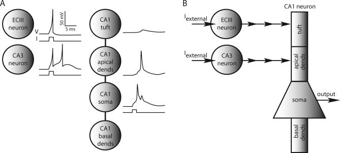
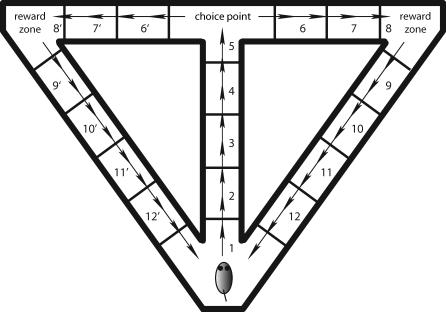
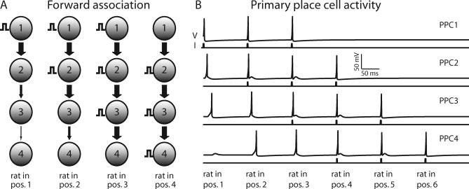
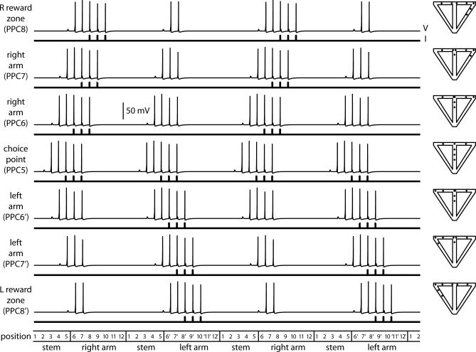
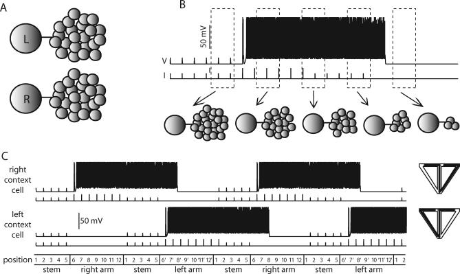
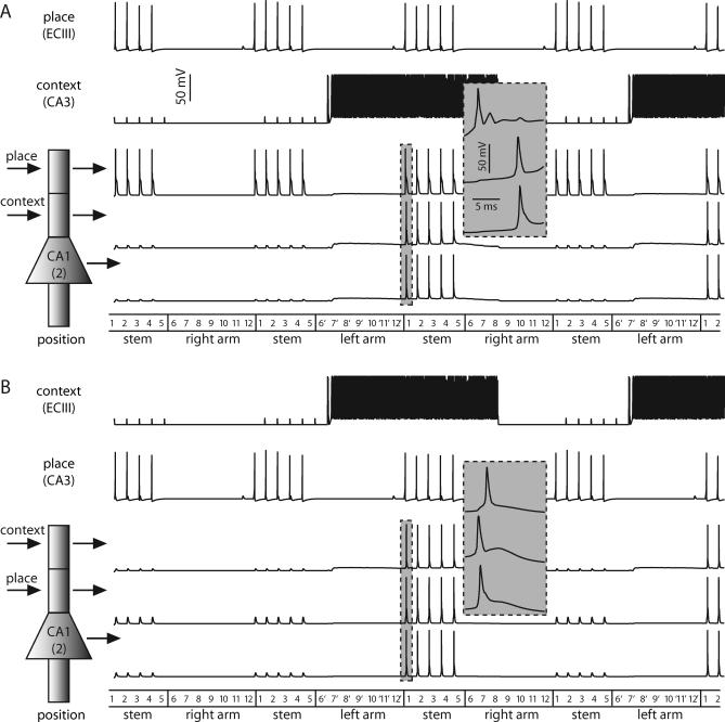
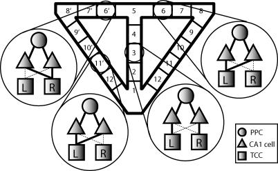
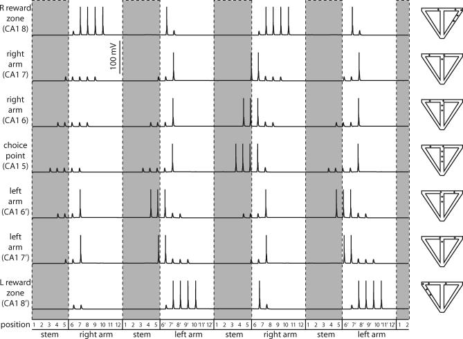
Recent advances in single-neuron biophysics have enhanced our understanding of information processing on the cellular level, but how the detailed properties of individual neurons give rise to large-scale behavior remains unclear. Here, we present a model of the hippocampal network based on observed biophysical properties of hippocampal and entorhinal cortical neurons. We assembled our model to simulate spatial alternation, a task that requires memory of the previous path through the environment for correct selection of the current path to a reward site. The convergence of inputs from entorhinal cortex and hippocampal region CA3 onto CA1 pyramidal cells make them potentially important for integrating information about place and temporal context on the network level. Our model shows how place and temporal context information might be combined in CA1 pyramidal neurons to give rise to splitter cells, which fire selectively based on a combination of place and temporal context. The model leads to a number of experimentally testable predictions that may lead to a better understanding of the biophysical basis of information processing in the hippocampus.


Conflict of interest statement

Competing interests. The authors have declared that no competing interests exist.

Figures

Figure 1
Figure 1. Elements of the Network Model
(A) ECIII and CA3 neurons are represented by single ECIII and CA3 nodes. Reduced model of a CA1 pyramidal neuron consists of four CA1 nodes electrically coupled together, representing the apical tuft, more-proximal apical dendrites (dends), soma, and basal dendrites. Shown are voltage responses of single, uncoupled ECIII, CA3, and CA1 neurons to 2-ms current injections of 200 pA, 200 pA, and 375 pA, respectively. The backpropagating action potential into the apical dendritic compartments of our CA1 pyramidal cell model shows that it has weakly excitable dendrites. (B) ECIII and CA3 cells receive external current inputs during the simulations. An ECIII cell provides input to the distal dendritic compartment of a CA1 cell, and a CA3 cell innervates its proximal dendritic compartment. Synaptic potentials are modeled as alpha functions, and if the voltage in the presynaptic cell exceeds −30 mV, an input is given to the postsynaptic cell.
Figure 2
Figure 2. The Virtual Environment
The virtual rat is confined to move through a T-maze with return arms. Although it moves in small steps, the maze is divided into larger positions, numbered 1–5 for positions on the stem, 6–12 for positions on the right, and 6′–12′ for corresponding positions on the left. The rat begins in position 1 at the base of the stem, moving up the stem to the choice point at the top of the stem. Virtual reward zones are in the right and left corners of the maze. The arrows denote a correct trajectory with the rat alternating between right and left turns at the choice point.
Figure 3
Figure 3. The Network of Primary Place Cells
(A) First column: when the rat enters position 1, PPC1 receives an external input. w factors are decreased (see text) so that the input propagates forward to PPC3, but the response in PPC4 is below spike threshold. Second column: when the rat enters position 2, all w factors are reset. PPCs 1 and 2 receive external inputs that elicit spiking in PPCs 3 and 4, but not PPC5. When the rat enters positions 3 and 4, external inputs are delivered and w factors are adjusted in a similar manner (remaining columns). (B) Time series plots for the primary place cells representing positions 1, 2, 3, and 4 on the maze. For each cell, the bottom trace is the input current and the top trace is the voltage response. PPCx gets external input at positions x, x + 1, and x + 2, and forward association input from positions x − 1 and x − 2. Therefore, a PPC spikes at most in five positions.
Figure 4
Figure 4. Forward Association from the Choice Point
Left: time-series plots for the PPCs representing the choice point and the three positions to the right and left of the choice point. For each cell, the bottom trace is the input current (200 pA) and the top trace is the voltage response. Activity spreads symmetrically from the choice point cell to the cells representing the right and left arms of the maze. Right: place fields for the cells depicted on the left. The dots represent spikes, showing where the animal was located when the cell fired.
Figure 5
Figure 5. The Network of Temporal Context Cells
(A) At first, the left (L) and right (R) TCCs are each connected to large recurrent networks of 22 cells each. (B) When the rat enters the right arm of the maze, the right TCC receives strong external input (200 pA), causing it to fire. With successive spiking, the right TCC can recruit a smaller and smaller portion of its network. The TCC continues to fire without external input only as long as it can recruit a recurrent network of sufficient strength. (C) Left: time-series plots for the right and left TCCs. Bottom traces are the input current, and top traces are the voltage response. Note that the magnitude of the input current increases (from 100 to 200 pA) as the rat moves from the stem into the arms of the maze. Right: context–place fields for the cells on the left. Note that the context–place fields are extremely broad.
Figure 6
Figure 6. Gating in the Reduced Model of a CA1 Pyramidal Neuron
(A) Shown are the cells representing position 2 of the maze. Here, the ECIII cell encodes place information and the CA3 cell represents temporal context. The CA1 cell only fires somatic spikes when the ECIII and CA3 inputs are coincident. As the rat enters the stem from the right arm, the subthreshold responses in the proximal apical dendrites and soma correspond to dendritic spikes that fail as they propagate forward. The gray inset shows the first set of CA1 spikes on an expanded time scale. (B) Same as above except the ECIII cell represents temporal context and the CA3 cell encodes raw place information. Although the somatic action potential profiles in (A) and (B) are roughly identical; in this case, the spike is initiated in the proximal apical dendritic compartment and propagates forward to the soma and backward to the apical tuft, as can be seen in the gray inset.
Figure 7
Figure 7. Network Wiring Diagram
Circles represent PPCs, triangles represent CA1 cells, squares represent TTCs cells, and lines indicate connections. The solid lines show robust connections that came about as a result of a presumed learning process, whereas the dashed lines suggest weak connections that have not been strengthened due to learning. Cells representing positions in the stem of the maze are connected as depicted for position 3: one CA1 cell for position 3 is connected to PPC3 and the right (R) TCC, whereas the other CA1 cell representing position 3 is connected to PPC3 and the left (L) TCC. Cells representing positions in the arms of the maze (except for positions on either side of the choice point, see below) are connected in the same way as the cells for position 11′. Both CA1 cells representing position 11′ are connected to PPC11′ and to the TCC representing the ipsilateral side of the maze, in this case the left TCC. The exception to this occurs in positions 6, 7, 6′, and 7′, which are wired as follows: both CA1 cells representing each position are connected to PPC representing that position and to the TCC representing the opposite side of the maze from which the position is located.
Figure 8
Figure 8. Firing Patterns of CA1 Cells near the Choice Point
Left: time-series data for the somata of CA1 neurons representing the choice point and the three positions to the right and left of the choice point. There are two CA1 cells associated with each location, as described in the text; only one cell for each location is shown. For the cell representing the choice point (CA1 5), the cell shown is the one associated with a right turn. Here, PPCs are taken to be ECIII cells, and TCCs to be CA3 cells, but when the representations are switched, the time series is essentially unchanged. Note that this figure includes the first time the rat goes through the maze, so it contains the initial transient where the final dynamics of all the neurons have not yet been established. Right: place fields for the somata of the cells on the left.
Figure 9
Figure 9. The CA1 Network
Raster plot showing spiking patterns for the entire CA1 network (including the initial transient). Cell number is plotted against position, and a vertical bar indicates a somatic spike when the rat is in a particular position. Cells in the stem are splitter cells: CA1 cells 1–5 R and 1–5 L fire only after right-turn and left-turn trials, respectively. The lines show how the rat can use the output of its CA1 cells to determine correct trajectories through the maze.

Similar articles

Cited by

References

    1. Wood ER, Dudchenko PA, Robitsek RJ, Eichenbaum H. Hippocampal neurons encode information about different types of memory episodes occurring in the same location. Neuron. 2000;27:623–633. - PubMed
    1. Hasselmo ME, Eichenbaum H. Hippocampal mechanisms for the context-dependent retrieval of episodes. Neural Netw. 2005;18:1172–1190. - PMC - PubMed
    1. Leutgeb S, Leutgeb JK, Moser MB, Moser EI. Place cells, spatial maps and the population code for memory. Curr Opin Neurobiol. 2005;15:738–746. - PubMed
    1. O'Keefe J, Burgess N. Dual phase and rate coding in hippocampal place cells: theoretical significance and relationship to entorhinal grid cells. Hippocampus. 2005;15:853–866. - PMC - PubMed
    1. Shapiro ML, Kennedy PJ, Ferbinteanu J. Representing episodes in the mammalian brain. Curr Opin Neurobiol. 2006;16:701–709. - PubMed

Publication types