

A cellular basis for Wolbachia recruitment to the host germline
- PMID: 18085821
- PMCID: PMC2134955
- DOI: 10.1371/journal.ppat.0030190
A cellular basis for Wolbachia recruitment to the host germline
Abstract
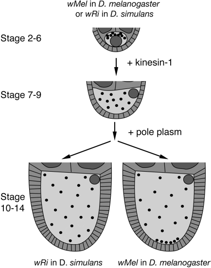
Wolbachia are among the most widespread intracellular bacteria, carried by thousands of metazoan species. The success of Wolbachia is due to efficient vertical transmission by the host maternal germline. Some Wolbachia strains concentrate at the posterior of host oocytes, which promotes Wolbachia incorporation into posterior germ cells during embryogenesis. The molecular basis for this localization strategy is unknown. Here we report that the wMel Wolbachia strain relies upon a two-step mechanism for its posterior localization in oogenesis. The microtubule motor protein kinesin-1 transports wMel toward the oocyte posterior, then pole plasm mediates wMel anchorage to the posterior cortex. Trans-infection tests demonstrate that factors intrinsic to Wolbachia are responsible for directing posterior Wolbachia localization in oogenesis. These findings indicate that Wolbachia can direct the cellular machinery of host oocytes to promote germline-based bacterial transmission. This study also suggests parallels between Wolbachia localization mechanisms and those used by other intracellular pathogens.
Conflict of interest statement
Figures
Similar articles
-
Wolbachia and host germline components compete for kinesin-mediated transport to the posterior pole of the Drosophila oocyte.PLoS Pathog. 2018 Aug 15;14(8):e1007216. doi: 10.1371/journal.ppat.1007216. eCollection 2018 Aug. PLoS Pathog. 2018. PMID: 30110391 Free PMC article.
-
Wolbachia utilizes host microtubules and Dynein for anterior localization in the Drosophila oocyte.PLoS Pathog. 2005 Oct;1(2):e14. doi: 10.1371/journal.ppat.0010014. Epub 2005 Oct 14. PLoS Pathog. 2005. PMID: 16228015 Free PMC article.
-
Distinct Wolbachia localization patterns in oocytes of diverse host species reveal multiple strategies of maternal transmission.Genetics. 2023 May 4;224(1):iyad038. doi: 10.1093/genetics/iyad038. Genetics. 2023. PMID: 36911919 Free PMC article.
-
The genetics and cell biology of Wolbachia-host interactions.Annu Rev Genet. 2008;42:683-707. doi: 10.1146/annurev.genet.41.110306.130354. Annu Rev Genet. 2008. PMID: 18713031 Review.
-
Impact of Wolbachia infection on Drosophila female germline stem cells.Curr Opin Insect Sci. 2020 Feb;37:8-15. doi: 10.1016/j.cois.2019.10.001. Epub 2019 Oct 17. Curr Opin Insect Sci. 2020. PMID: 31726321 Review.
Cited by
-
Pervasive effects of Wolbachia on host activity.Biol Lett. 2021 May;17(5):20210052. doi: 10.1098/rsbl.2021.0052. Epub 2021 May 5. Biol Lett. 2021. PMID: 33947218 Free PMC article.
-
Wolbachia and host germline components compete for kinesin-mediated transport to the posterior pole of the Drosophila oocyte.PLoS Pathog. 2018 Aug 15;14(8):e1007216. doi: 10.1371/journal.ppat.1007216. eCollection 2018 Aug. PLoS Pathog. 2018. PMID: 30110391 Free PMC article.
-
The Maternal Effect Gene Wds Controls Wolbachia Titer in Nasonia.Curr Biol. 2018 Jun 4;28(11):1692-1702.e6. doi: 10.1016/j.cub.2018.04.010. Epub 2018 May 17. Curr Biol. 2018. PMID: 29779872 Free PMC article.
-
Effects of Wolbachia infection and ovarian tumor mutations on Sex-lethal germline functioning in Drosophila.Genetics. 2009 Apr;181(4):1291-301. doi: 10.1534/genetics.108.099374. Epub 2009 Jan 26. Genetics. 2009. PMID: 19171941 Free PMC article.
-
Large-Scale Identification of Wolbachia pipientis Effectors.Genome Biol Evol. 2017 Jul 1;9(7):1925-1937. doi: 10.1093/gbe/evx139. Genome Biol Evol. 2017. PMID: 28854601 Free PMC article.
References
-
- Tram U, Ferree PM, Sullivan W. Identification of Wolbachia--host interacting factors through cytological analysis. Microbes Infect. 2003;5:999–1011. - PubMed
-
- Stouthamer R, Breeuwer JA, Hurst GD. Wolbachia pipientis: microbial manipulator of arthropod reproduction. Annu Rev Microbiol. 1999;53:71–102. - PubMed
-
- Hise AG, Gillette-Ferguson I, Pearlman E. The role of endosymbiotic Wolbachia bacteria in filarial disease. Cell Microbiol. 2004;6:97–104. - PubMed
-
- Foster J, Ganatra M, Kamal I, Ware J, Makarova K, et al. The Wolbachia genome of Brugia malayi: endosymbiont evolution within a human pathogenic nematode. PLoS Biol. 2005;3:e121. doi: 10.1371/journal.pbio.0030121. - DOI - PMC - PubMed
-
- Saint Andre A, Blackwell NM, Hall LR, Hoerauf A, Brattig NW, et al. The role of endosymbiotic Wolbachia bacteria in the pathogenesis of river blindness. Science. 2002;295:1892–1895. - PubMed
Publication types
MeSH terms
Substances
Grants and funding
LinkOut - more resources
Full Text Sources
Other Literature Sources
Molecular Biology Databases
