. 2007 Dec;3(12):e191.
doi: 10.1371/journal.ppat.0030191.

Co-circulation and evolution of polioviruses and species C enteroviruses in a district of Madagascar

Affiliations

Co-circulation and evolution of polioviruses and species C enteroviruses in a district of Madagascar

Mala Rakoto-Andrianarivelo et al. PLoS Pathog. 2007 Dec.

Abstract

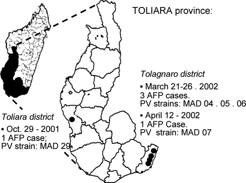
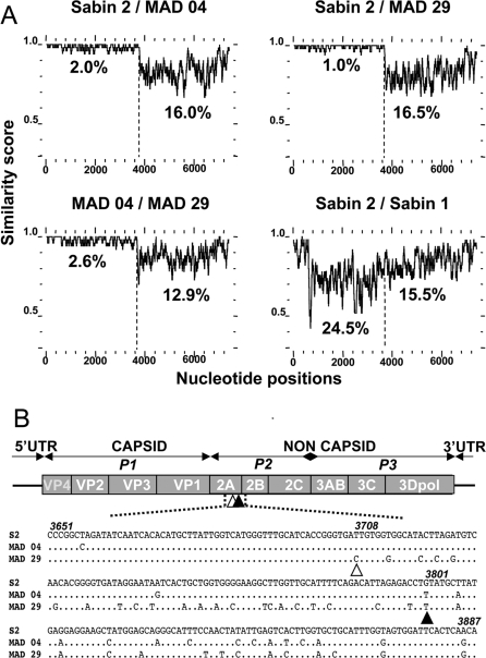
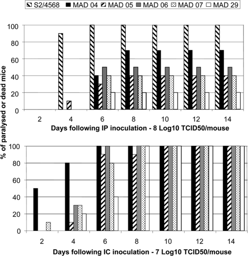
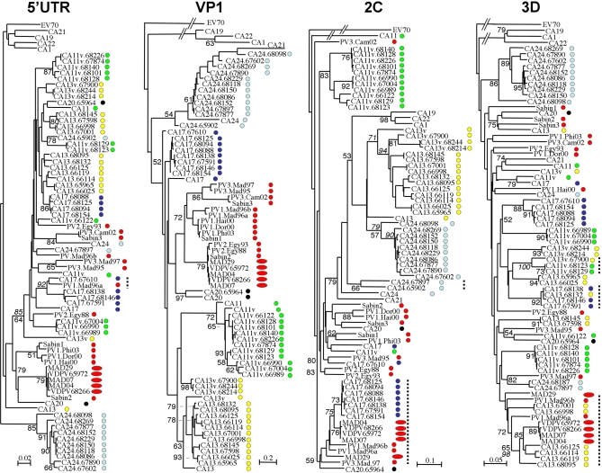
Between October 2001 and April 2002, five cases of acute flaccid paralysis (AFP) associated with type 2 vaccine-derived polioviruses (VDPVs) were reported in the southern province of the Republic of Madagascar. To determine viral factors that favor the emergence of these pathogenic VDPVs, we analyzed in detail their genomic and phenotypic characteristics and compared them with co-circulating enteroviruses. These VDPVs appeared to belong to two independent recombinant lineages with sequences from the type 2 strain of the oral poliovaccine (OPV) in the 5'-half of the genome and sequences derived from unidentified species C enteroviruses (HEV-C) in the 3'-half. VDPV strains showed characteristics similar to those of wild neurovirulent viruses including neurovirulence in poliovirus-receptor transgenic mice. We looked for other VDPVs and for circulating enteroviruses in 316 stools collected from healthy children living in the small area where most of the AFP cases occurred. We found vaccine PVs, two VDPVs similar to those found in AFP cases, some echoviruses, and above all, many serotypes of coxsackie A viruses belonging to HEV-C, with substantial genetic diversity. Several coxsackie viruses A17 and A13 carried nucleotide sequences closely related to the 2C and the 3D(pol) coding regions of the VDPVs, respectively. There was also evidence of multiple genetic recombination events among the HEV-C resulting in numerous recombinant genotypes. This indicates that co-circulation of HEV-C and OPV strains is associated with evolution by recombination, resulting in unexpectedly extensive viral diversity in small human populations in some tropical regions. This probably contributed to the emergence of recombinant VDPVs. These findings give further insight into viral ecosystems and the evolutionary processes that shape viral biodiversity.


Conflict of interest statement

Competing interests. The authors have declared that no competing interests exist.

Figures

Figure 1
Figure 1. Map of the Toliara Province in Madagascar
This map indicates the location of the two districts where the poliomyelitis outbreaks occurred. Names of the PV strains isolated from the patients with AFP are indicated.
Figure 2
Figure 2. Genomic Features of the Poliovirus Isolates
(A) Plots of similarity between a set of aligned genomes (Sabin 2, MAD 04, MAD 29, and Sabin 1), using a 50-nt sliding window. Approximate nucleotide positions in the poliovirus genome are indicated. Average nucleotide identities between the 5′ vaccine part and the 3′ non-vaccine part of the recombinant genomes are shown. (B) Sites of the recombination junctions in the recombinant VDPV genomes. The genetic organization of the PV genome is shown, including the 5′- and 3′-UTR. Genomic regions P1 to P3 encoding viral proteins (VP4 to 3Dpol) are indicated. Open and closed triangles indicate the approximate sites of the recombination junctions of the MAD 29 and MAD 04 recombinant genomes, respectively. Both junctions are located at the 3′ end of the protease 2A genomic region. MAD 04 and MAD 29 sequences (nt 3651–3890) on both sides of the recombination junctions are given and compared with Sabin 2 sequences (S2). Nucleotide numbering according to Sabin 2 sequences is given in italics.
Figure 3
Figure 3. Neurovirulence of VDPVs in Transgenic PVR-Tg Mice
A given dose of virus was used to inoculate intracerebrally (IC) or intraperitoneally (IP) groups of PVR-Tg mice expressing the human poliovirus receptor (8–10 animals per virus). Animals were checked daily for 14 d following inoculation for paralysis and death. Percentages of affected mice following inoculation of viruses MAD 04 to MAD 07, MAD 29, and of a positive neurovirulent control virus S2/4568 are given. Strain S2/4568 was used only for IP inoculation. IP or IC inoculation of the vaccine strain Sabin 2 did not induce disease in animals.
Figure 4
Figure 4. Phylogenetic Trees Depicting Genetic Relationships between Nucleotide Sequences of Enteroviruses of the HEV-C Species
These neighbor-joining trees were based on nucleotide sequence alignments of part of the 5′ UTR, the C-terminal third of the VP1-coding regions, part of the 2C-coding regions, and of the 3Dpol regions. Branch lengths were calculated using PUZZLE and the Hasegawa, Kishino, and Yano (HKY) model of substitution [63]. The genetic distance is indicated (bar). Numbers at nodes correspond to the percentage of 25,000 puzzle steps supporting the distal cluster; numbers in italics representing the percentage of 1,000 bootstrap pseudoreplicates obtained with the DNADist/Neighbor and SEQBOOT programs of PHYLIP [65] are shown in several cases. Sequences of some of the last wild PV strains (PV3.Mad95, PV1.Mad96a, PV1.Mad96b, and PV3.Mad97) isolated in Madagascar (1995–1997) were used in addition to HEV-C prototype strains (CA1 to CA24). Selected PV strains isolated during the last poliomyelitis outbreak in Finland (PV3.Fin84) and during the recent VDPV outbreaks in Egypt (PV2.Egy93; PV2.Egy88), Haiti (PV1.Hai00), Dominican Republic (PV1.Dor00), and the Philippines (PV1.Phi01) are included. Sequences of the recombinant type 3 PV isolated from a poliomyelitis case in Cambodia in 2002 (PV3.Cam02) were also used. Nucleotide sequences of echovirus 70 (EV70) were used as outgroup. Identifiers of the HEV-C coxsackie A viruses isolated from healthy children in 2002 in Madagascar consist of the given serotype as assessed from partial sequencing [33] and the laboratory number. New VDPV isolates from healthy children are indicated with their laboratory numbers. Spots of different colors differentiate HEV-C serotypes and polioviruses (red). VDPVs from Madagascar are indicated by oblong spots. According to the Eight Report of the ICTV [56], former CA15 and CA18 are considered as antigenic variants of CA11 and CA13, and are now named CA11v and CA13v, respectively. All field isolates from Madagascar, except isolates CA24.65902 and CA24.68187, were sequenced in the four regions. Vertical dotted lines indicate groups of isolates of particular interest that are discussed in the text.

Similar articles

Cited by

References

    1. Pallansch M, Roos R. Enteroviruses: polioviruses, coxsackieviruses, echoviruses, and newer enteroviruses. In: Knipe D, Howley P, editors. Fields virology. Philadelphia: Lippincott Williams and Wilkins; 2001. pp. 723–775.
    1. Anonymous. Progress toward interruption of wild poliovirus transmission worldwide, January 2006–May 2007. Morb Mortal Wkly Rep. 2007;56:682–685. - PubMed
    1. Minor PD. The molecular biology of poliovaccines. J Gen Virol. 1992;73:3065–3077. - PubMed
    1. Dahourou G, Guillot S, Le Gall O, Crainic R. Genetic recombination in wild-type poliovirus. J Gen Virol. 2002;83:3103–3110. - PubMed
    1. Liu HM, Zheng DP, Zhang LB, Oberste MS, Kew OM, et al. Serial recombination during circulation of type 1 wild-vaccine recombinant polioviruses in China. J Virol. 2003;77:10994–11005. - PMC - PubMed

Publication types

MeSH terms