

Hemophagocytic macrophages harbor Salmonella enterica during persistent infection
- PMID: 18085823
- PMCID: PMC2134957
- DOI: 10.1371/journal.ppat.0030193
Hemophagocytic macrophages harbor Salmonella enterica during persistent infection
Abstract
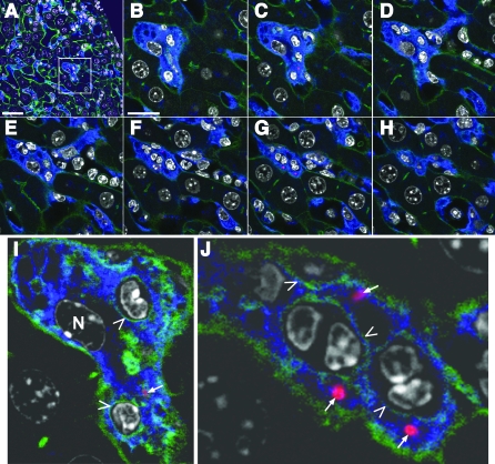
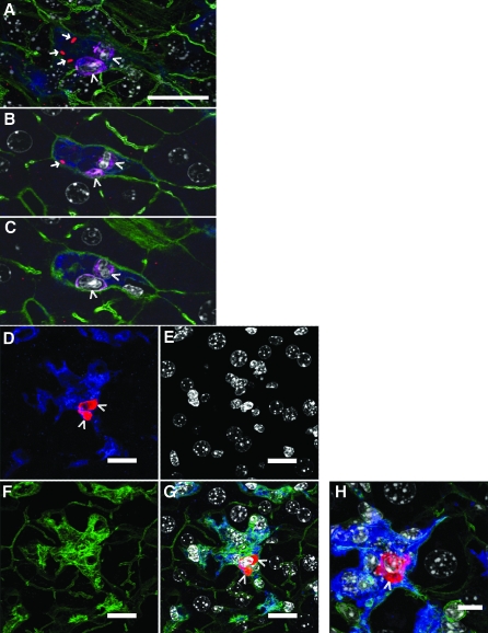
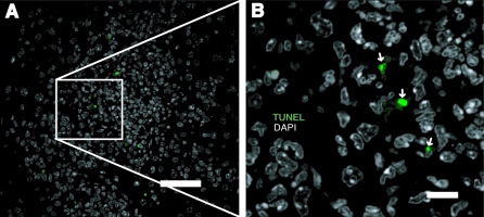
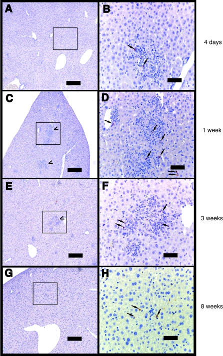
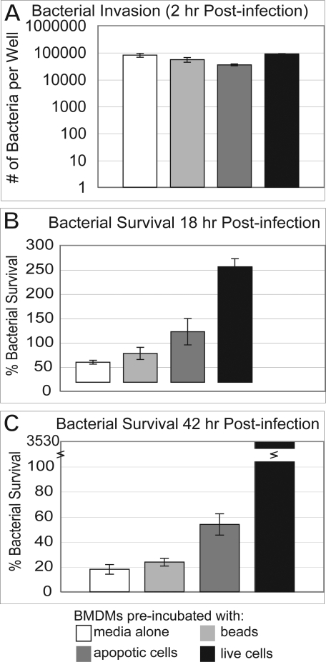
Salmonella enterica subspecies can establish persistent, systemic infections in mammals, including human typhoid fever. Persistent S. enterica disease is characterized by an initial acute infection that develops into an asymptomatic chronic infection. During both the acute and persistent stages, the bacteria generally reside within professional phagocytes, usually macrophages. It is unclear how salmonellae can survive within macrophages, cells that evolved, in part, to destroy pathogens. Evidence is presented that during the establishment of persistent murine infection, macrophages that contain S. enterica serotype Typhimurium are hemophagocytic. Hemophagocytic macrophages are characterized by the ingestion of non-apoptotic cells of the hematopoietic lineage and are a clinical marker of typhoid fever as well as certain other infectious and genetic diseases. Cell culture assays were developed to evaluate bacterial survival in hemophagocytic macrophages. S. Typhimurium preferentially replicated in macrophages that pre-phagocytosed viable cells, but the bacteria were killed in macrophages that pre-phagocytosed beads or dead cells. These data suggest that during persistent infection hemophagocytic macrophages may provide S. Typhimurium with a survival niche.
Conflict of interest statement
Figures
Similar articles
-
Salmonella enterica infection stimulates macrophages to hemophagocytose.mBio. 2014 Dec 9;5(6):e02211. doi: 10.1128/mBio.02211-14. mBio. 2014. PMID: 25491357 Free PMC article.
-
A comprehensive study of the contribution of Salmonella enterica serovar Typhimurium SPI2 effectors to bacterial colonization, survival, and replication in typhoid fever, macrophage, and epithelial cell infection models.Virulence. 2011 May-Jun;2(3):208-16. doi: 10.4161/viru.2.3.15894. Epub 2011 May 1. Virulence. 2011. PMID: 21540636 Free PMC article.
-
Production of Murine Macrophages from Hoxb8-Immortalized Myeloblasts: Utility and Use in the Context of Salmonella Infection.Methods Mol Biol. 2021;2182:117-126. doi: 10.1007/978-1-0716-0791-6_11. Methods Mol Biol. 2021. PMID: 32894491
-
The role of host cell death in Salmonella infections.Curr Top Microbiol Immunol. 2005;289:131-50. doi: 10.1007/3-540-27320-4_6. Curr Top Microbiol Immunol. 2005. PMID: 15791954 Review.
-
Intracellular microbes and haemophagocytosis.Cell Microbiol. 2008 Nov;10(11):2151-8. doi: 10.1111/j.1462-5822.2008.01192.x. Epub 2008 Jun 23. Cell Microbiol. 2008. PMID: 18616693 Free PMC article. Review.
Cited by
-
Hemophagocytic macrophages in murine typhoid fever have an anti-inflammatory phenotype.Infect Immun. 2012 Oct;80(10):3642-9. doi: 10.1128/IAI.00656-12. Epub 2012 Aug 6. Infect Immun. 2012. PMID: 22868497 Free PMC article.
-
Dissemination of persistent intestinal bacteria via the mesenteric lymph nodes causes typhoid relapse.Infect Immun. 2011 Apr;79(4):1479-88. doi: 10.1128/IAI.01033-10. Epub 2011 Jan 24. Infect Immun. 2011. PMID: 21263018 Free PMC article.
-
Shigella Serotypes Associated With Carriage in Humans Establish Persistent Infection in Zebrafish.J Infect Dis. 2023 Oct 18;228(8):1108-1118. doi: 10.1093/infdis/jiad326. J Infect Dis. 2023. PMID: 37556724 Free PMC article.
-
Iron and infection.Int J Hematol. 2018 Jan;107(1):7-15. doi: 10.1007/s12185-017-2366-2. Epub 2017 Nov 16. Int J Hematol. 2018. PMID: 29147843 Review.
-
Salmonella enterica replication in hemophagocytic macrophages requires two type three secretion systems.Infect Immun. 2010 Aug;78(8):3369-77. doi: 10.1128/IAI.00292-10. Epub 2010 Jun 1. Infect Immun. 2010. PMID: 20515933 Free PMC article.
References
-
- Parry CM, Hien TT, Dougan G, White NJ, Farrar JJ. Typhoid fever. N Engl J Med. 2002;347:1770–1782. - PubMed
-
- Plant JE, Blackwell JM, O'Brien AD, Bradley DJ, Glynn AA. Are the Lsh and Ity disease resistance genes at one locus on mouse chromosome 1? Nature. 1982;297:510–511. - PubMed
-
- Monack DM, Mueller A, Falkow S. Persistent bacterial infections: the interface of the pathogen and the host immune system. Nat Rev Microbiol. 2004;2:747–765. - PubMed
-
- Coburn B, Grassl GA, Finlay BB. Salmonella, the host and disease: a brief review. Immunol Cell Biol. 2007;85:112–118. - PubMed
Publication types
MeSH terms
Substances
Grants and funding
LinkOut - more resources
Full Text Sources
Other Literature Sources
Medical
