

Melatonin attenuates calpain upregulation, axonal damage and neuronal death in spinal cord injury in rats
- PMID: 18086148
- PMCID: PMC2613550
- DOI: 10.1111/j.1600-079X.2007.00534.x
Melatonin attenuates calpain upregulation, axonal damage and neuronal death in spinal cord injury in rats
Abstract
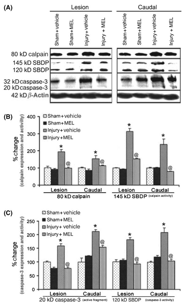
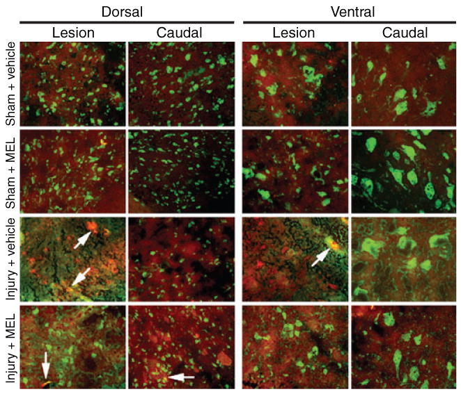
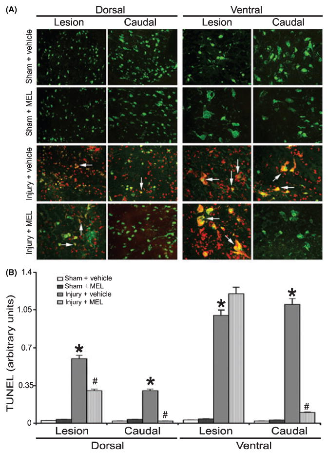
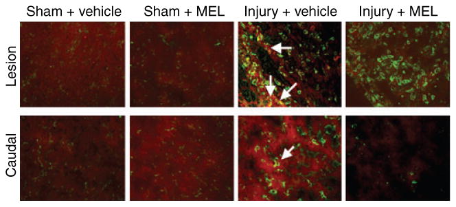
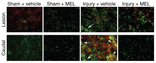
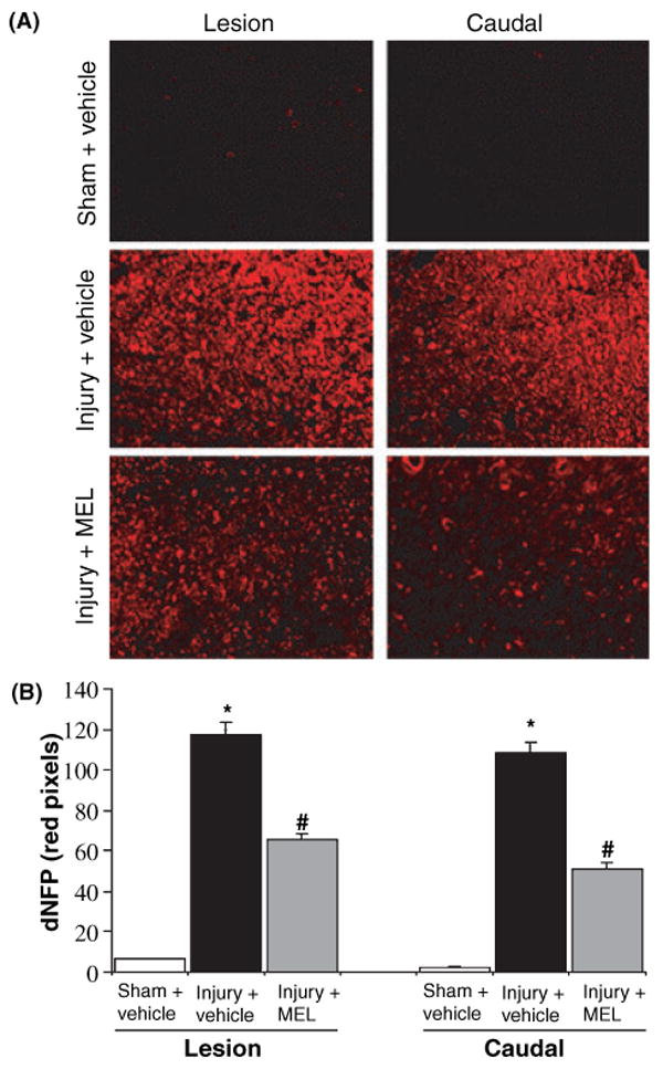
Multiple investigations in vivo have shown that melatonin (MEL) has a neuroprotective effect in the treatment of spinal cord injury (SCI). This study investigates the role of MEL as an intervening agent for ameliorating Ca(2+)-mediated events, including activation of calpain, following its administration to rats sustaining experimental SCI. Calpain, a Ca(2+)-dependent neutral protease, is known to be involved in the pathogenesis of SCI. Rats were injured using a standard weight-drop method that induced a moderately severe injury (40 g.cm force) at T10. Sham controls received laminectomy only. Injured animals were given either 45 mg/kg MEL or vehicle at 15 min post-injury by intraperitoneal injection. At 48 hr post-injury, spinal cord (SC) samples were collected. Immunofluorescent labelings were used to identify calpain expression in specific cell types, such as neurons, glia, or macrophages. Combination of terminal deoxynucleotidyl transferase (TdT)-mediated dUTP nick-end labeling (TUNEL) and double immunofluorescent labelings was used to identify apoptosis in specific cells in the SC. The effect of MEL on axonal damage was also investigated using antibody specific for dephosphorylated neurofilament protein (dNFP). Treatment of SCI animals with MEL attenuated calpain expression, inflammation, axonal damage (dNFP), and neuronal death, indicating that MEL provided neuroprotective effect in SCI. Further, expression and activity of calpain and caspse-3 were examined by Western blotting. The results indicated a significant decrease in expression and activity of calpain and caspse-3 in SCI animals after treatment with MEL. Taken together, this study strongly suggested that MEL could be an effective neuroprotective agent for treatment of SCI.
Figures
Similar articles
-
Direct evidence for calpain involvement in apoptotic death of neurons in spinal cord injury in rats and neuroprotection with calpain inhibitor.Neurochem Res. 2007 Dec;32(12):2210-6. doi: 10.1007/s11064-007-9433-7. Epub 2007 Aug 4. Neurochem Res. 2007. PMID: 17676387
-
Early induction of secondary injury factors causing activation of calpain and mitochondria-mediated neuronal apoptosis following spinal cord injury in rats.J Neurosci Res. 2003 Jul 1;73(1):95-104. doi: 10.1002/jnr.10607. J Neurosci Res. 2003. PMID: 12815713
-
Estrogen treatment of spinal cord injury attenuates calpain activation and apoptosis.J Neurosci Res. 2006 Oct;84(5):1064-75. doi: 10.1002/jnr.21016. J Neurosci Res. 2006. PMID: 16902996
-
Inhibition of cysteine proteases in acute and chronic spinal cord injury.Neurotherapeutics. 2011 Apr;8(2):180-6. doi: 10.1007/s13311-011-0037-1. Neurotherapeutics. 2011. PMID: 21373949 Free PMC article. Review.
-
Neuroprotective efficacy of estrogen in experimental spinal cord injury in rats.Ann N Y Acad Sci. 2010 Jun;1199:90-4. doi: 10.1111/j.1749-6632.2009.05357.x. Ann N Y Acad Sci. 2010. PMID: 20633113 Free PMC article. Review.
Cited by
-
Premarin Reduces Neurodegeneration and Promotes Improvement of Function in an Animal Model of Spinal Cord Injury.Int J Mol Sci. 2022 Feb 21;23(4):2384. doi: 10.3390/ijms23042384. Int J Mol Sci. 2022. PMID: 35216504 Free PMC article.
-
Reduction in traumatic brain injury-induced oxidative stress, apoptosis, and calcium entry in rat hippocampus by melatonin: Possible involvement of TRPM2 channels.Metab Brain Dis. 2015 Feb;30(1):223-31. doi: 10.1007/s11011-014-9623-3. Epub 2014 Oct 23. Metab Brain Dis. 2015. PMID: 25339252
-
Triad1 Promotes the Inflammatory Response and Neuronal Apoptosis to Aggravate Acute Spinal Cord Injury in Rats.Comput Math Methods Med. 2022 Jul 20;2022:2025756. doi: 10.1155/2022/2025756. eCollection 2022. Comput Math Methods Med. 2022. Retraction in: Comput Math Methods Med. 2023 Jul 12;2023:9754812. doi: 10.1155/2023/9754812. PMID: 35912142 Free PMC article. Retracted.
-
Neurotrauma and mesenchymal stem cells treatment: From experimental studies to clinical trials.World J Stem Cells. 2014 Apr 26;6(2):179-94. doi: 10.4252/wjsc.v6.i2.179. World J Stem Cells. 2014. PMID: 24772245 Free PMC article. Review.
-
Melatonin treatment protects against acute spinal cord injury-induced disruption of blood spinal cord barrier in mice.J Mol Neurosci. 2014 Dec;54(4):714-22. doi: 10.1007/s12031-014-0430-4. Epub 2014 Oct 11. J Mol Neurosci. 2014. PMID: 25303856
References
-
- National Science Statistical Center. National Spinal Cord Injury Center; Birmingham, AL: 2006. Spinal cord injury: Facts and FIgures at a glance.
-
- Ray SK, Hogan EL, Banik NL. Calpain in the pathophysiology of spinal cord injury: neuroprotection with calpain inhibitors. Brain Res Brain Res Rev. 2003;42:169–185. - PubMed
-
- Ray SK, Banik NL. Chaplain and its involvement in the path physiology of CNS injuries and diseases: therapeutic potential of chaplain inhibitors for prevention of neurodegeneration. Curr Drug Targets CNS Neurol Disord. 2003;2:173–189. - PubMed
-
- Dumont RJ, Verma S, Okonkwo DO, et al. Acute spinal cord injury, part II: contemporary pharmacotherapy. Clin Neuropharmacol. 2001;24:265–279. - PubMed
-
- Zimmerman UJ, Boring L, Pak JH, et al. The calpain small subunit gene is essential: its inactivation results in embryonic lethality. IUBMB Life. 2000;50:63–68. - PubMed
Publication types
MeSH terms
Substances
Grants and funding
- NS-41088/NS/NINDS NIH HHS/United States
- NS-38146/NS/NINDS NIH HHS/United States
- R01 NS031622/NS/NINDS NIH HHS/United States
- R01 NS045967/NS/NINDS NIH HHS/United States
- CA-91460/CA/NCI NIH HHS/United States
- VM08716/PHS HHS/United States
- NS-45967/NS/NINDS NIH HHS/United States
- C06 RR015455/RR/NCRR NIH HHS/United States
- R01 CA091460/CA/NCI NIH HHS/United States
- T32 GM008716/GM/NIGMS NIH HHS/United States
- R01 NS041088/NS/NINDS NIH HHS/United States
- R01 NS057811/NS/NINDS NIH HHS/United States
- NS-57811/NS/NINDS NIH HHS/United States
- R01 NS-316222/NS/NINDS NIH HHS/United States
- R01 NS038146/NS/NINDS NIH HHS/United States
LinkOut - more resources
Full Text Sources
Medical
Research Materials
Miscellaneous
