Review
. 2007;212(9-10):771-84.
doi: 10.1016/j.imbio.2007.09.018. Epub 2007 Nov 28.

Emerging themes in IFN-gamma-induced macrophage immunity by the p47 and p65 GTPase families

Affiliations
Review

Emerging themes in IFN-gamma-induced macrophage immunity by the p47 and p65 GTPase families

Avinash R Shenoy et al. Immunobiology. 2007.

Abstract

Vertebrates have evolved complex immune specificity repertoires beyond the primordial components found in lower multi-cellular organisms to combat microbial infections. The type II interferon (IFN-gamma) pathway represents one such system, bridging innate and acquired immunity and providing host protection in a cell-autonomous manner. Recent large-scale transcriptome analyses of IFN-gamma-dependent gene expression in effector cells such as macrophages have highlighted the prominence of two families of GTPases -- p47 IRGs and p65 GBPs -- that are now beginning to emerge as major determinants of antimicrobial resistance. Here we discuss the recent clarification of known family members, their cellular biochemistry and host defense functions as a means to understanding the complex innate immune response engendered in higher vertebrates such as humans and mice.


Figures
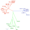
Fig. 1
Fig. 1
Phylogenetic analyses of p47 IRGs and p65 GBPs. G-domains of p47 IRGs, p65 GBPs and other representative GTPases were first aligned separately using ProbCons (Do et al., 2005) followed by two step profiles using TCoffee (Notredame et al., 2000) which generated the best alignment of this divergent group of proteins. The neighbor-joining tree shown was based on this alignment using the Molecular Evolutionary Genetics Analysis package (version 3.1; Kumar et al., 2004). The p47 IRGs separate into distinct sub-families, particularly the N-and C-terminal GTPase domains (indicated as G1 and G2 respectively) of L-Irg1-3 that form two sub-clusters. Human IRGM lies nearest to the cluster containing mouse Lrg-47/Irgm1, Gtpi/Irgm2 and Igtp/Irgm3. Note the groupings within the GBP family indicating a closer relation between genes in this family. Scale bar designates number of substitutions per site.
Fig. 2
Fig. 2
Genomic organization of human and mouse p47 IRGs and p65 GBPs. (A) Schematic representation of the human genomic regions containing p47 IRG and p65 GBP genes on chromosomes 1, 5 and 19. Arrows represent 5′–3′ direction of mRNA transcripts and boxes represent the canonical Iigp1/Irga6-like GTPase domain (Pfam accession PF05049). Small green boxes are presumed GBP pseudogenes. Red boxes represent genes unrelated to the GBP family. (B) Schematic representation of the mouse genomic regions containing multiple p47 Irg and p65 Gbp genes on chromosomes 3, 5, 7, 11 and 18. Three longer genes (labeled L-Irg1-3) may contain two tandem GTPase domains previously treated as individual proteins (Bekpen et al., 2005). The N-terminal GTPase domain of L-Irg1 is identical to Irgb5, a genomic duplication that has been pointed out earlier (Bekpen et al., 2005). Blue boxes indicate presumed IRG pseudogenes (Irgb7, Irga1, Irga5 and Irga8) and red boxes represent genes unrelated to the IRG family. Irg11 is a new IRG gene upstream of Iigp1/Irgb6. For the p65 Gbp family, new genes have been labeled as Gbp8-13. The gene nomenclature endorsed by HUGO is shown on the top, with NCBI GeneID numbers below.
Fig. 3
Fig. 3
Structures and sequence relationship within the p47 IRGs and p65 GBPs. (A) A computer rendering of full-length dimeric Iigp1/Irgb6 (pdb code 1tq2) crystallized in the presence of phosphoaminophosphonic acid-guanylate ester (GNP; shown in red stick representation). Subunits in the dimer are colored in shades of blue and green. For the sake of clarity, N- and C-terminal helical domains and the G-domain are shaded and marked as indicated for the protomer on the left. The axis relates the protomers by 2-fold non-crystallographic symmetry. The αK region responsible for membrane targeting in Lrg47/Irgm1 is indicated. (B) A model of monomeric full-length hGBP1 (pdb code 1f5n) shaded to represent its subdomains. The elongated nature of hGBP1 versus Iigp1/Irga6 is apparent. The active site nucleotide (GNP; red stick model) was imported from the hGBP1 G-domain crystal structure (pdb code 2bc9). (C) Conserved motifs from an alignment of G-domains of p47 IRGs and p65 GBPs (see legend to Fig. 1) were identified using BLOCKS (Henikoff and Henikoff, 1991), and LOGO (Schneider and Stephens, 1990) representations of these are shown in the panel on top. The bottom panel shows a closer view of the G-domain of Iigp1/Irga6 (1tq2) and hGBP1 (2bc9). Positions of the conserved GTPase motifs shown in the upper panel are depicted with the rest of the protein drawn as ribbons. The divergent switch-I region is shown in red and orange for the IRGs and GBPs, respectively. GBPs lack a G5 motif but instead have a ‘guanine cap’ that closes the nucleotide binding pocket as shown in blue.
Fig. 4
Fig. 4
IFN-inducible transcription factor-binding sites within p47 IRG and p65 GBP promoters. Results from earlier studies of known genes are summarized schematically (Bekpen et al., 2005; Olszewski et al., 2006). Analyses of newly identified members were carried out using P-match (Chekmenev et al., 2005), and results for GAS (interferon-gamma activated site) or ISRE (interferon stimulated response elements) are shown.
Fig. 5
Fig. 5
Recruitment of endogenous Lrg-47/Irgm1 to the bacterial phagosome. Upper panel depicts single-channel and merged (overlay) confocal images of Lrg-47/Irgm1 on the mycobacterial phagosome (M. bovis BCG) in IFN-γ-activated RAW264.7 murine macrophages. Antigens were detected using goat anti-Lrg-47 peptide (A19) pAb coupled to Alexa-488 with rabbit anti-PPD pAb-linked Alexa 594. Lower panel demonstrates early recruitment (10–15 min post-uptake) of Lrg-47/Irgm1 to the L. monocytogenes vacuole in IFN-γ-activated mouse macrophages. Rabbit anti-L. monocytogenes (ATCC strain 43251) pAb was used for bacterial visualization. Original magnification, 100×. Courtesy of T. Matsuzawa.

References

    1. Anderson SL, Carton JM, Lou J, Xing L, Rubin BY. Interferon-induced guanylate binding protein-1 (GBP-1) mediates an antiviral effect against vesicular stomatitis virus and encephalomyocarditis virus. Virology. 1999;256:8–14. - PubMed
    1. Bekpen C, Hunn JP, Rohde C, Parvanova I, Guethlein L, Dunn DM, Glowalla E, Leptin M, Howard JC. The interferon-inducible p47 (IRG) GTPases in vertebrates: loss of the cell autonomous resistance mechanism in the human lineage. Genome Biol. 2005;6:R92. - PMC - PubMed
    1. Bernstein-Hanley I, Coers J, Balsara ZR, Taylor GA, Starnbach MN, Dietrich WF. The p47 GTPases Igtp and Irgb10 map to the Chlamydia trachomatis susceptibility locus Ctrq-3 and mediate cellular resistance in mice. Proc Natl Acad Sci USA. 2006;103:14092–14097. - PMC - PubMed
    1. Boehm U, Guethlein L, Klamp T, Ozbek K, Schaub A, Futterer A, Pfeffer K, Howard JC. Two families of GTPases dominate the complex cellular response to IFN-gamma. J Immunol. 1998;161:6715–6723. - PubMed
    1. Briken V, Ruffner H, Schultz U, Schwarz A, Reis LF, Strehlow I, Decker T, Staeheli P. Interferon regulatory factor 1 is required for mouse Gbp gene activation by gamma interferon. Mol Cell Biol. 1995;15:975–982. - PMC - PubMed

Publication types

LinkOut - more resources